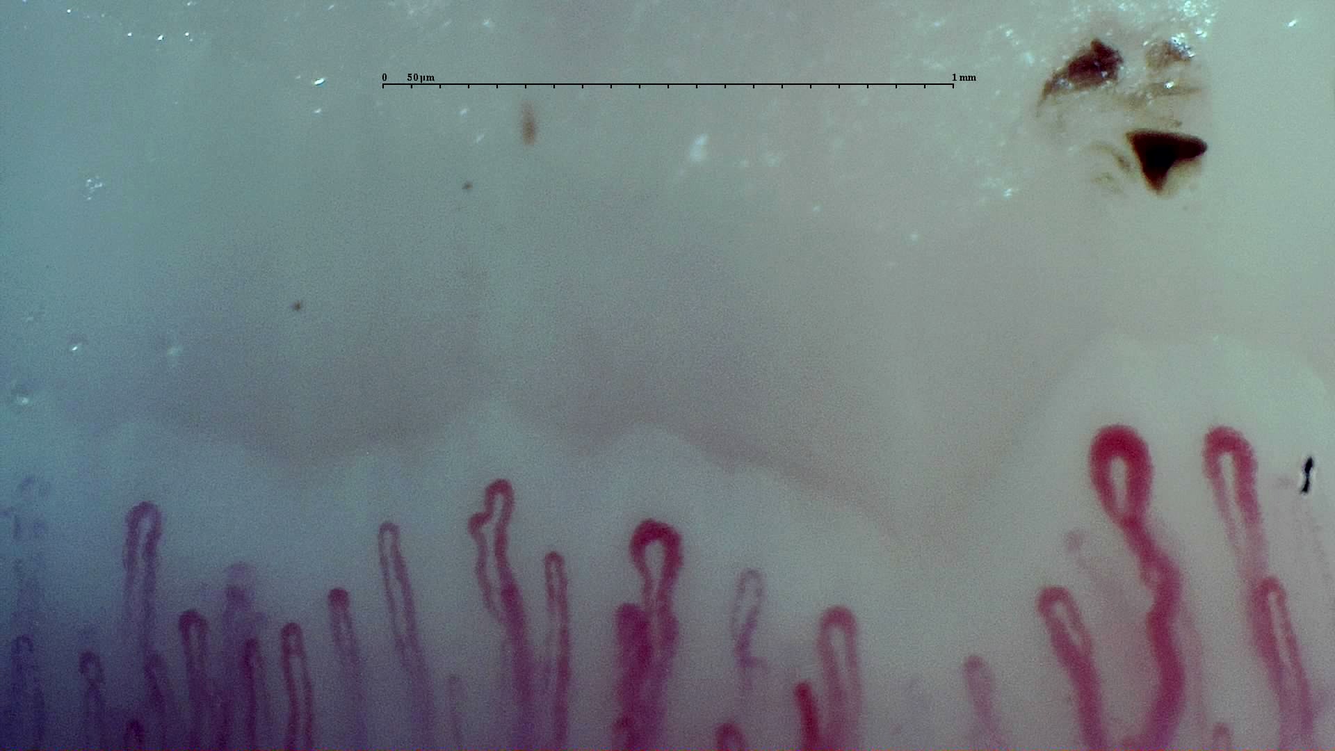
 Early scleroderma pattern: selected field
Selected field from the early-pattern reference study, with enlarged loops among a relatively preserved capillary row. The subtype belongs to the complete examination.
View the complete studyHow to assess and measure
Examine several assessable fields across fingers, confirm giant dimensions with calibration and document hemorrhages, density and organization. Preserved density does not exclude an early pattern when characteristic giants and hemorrhages are present. Use the complete reference study below to check distribution beyond a single selected image.
Interpretation and related diseases
Integrate the pattern with Raynaud’s history, examination and autoantibodies when assessing possible systemic sclerosis. An isolated dilated loop or traumatic hemorrhage is insufficient. Scleroderma-like changes can occur in other connective tissue diseases; the pattern label should not replace clinical differential diagnosis.
Read the detailed clinical chapterCommon mistakes
- Calling a nonspecific dilation a giant capillary.
- Excluding relevant microangiopathy just because density is preserved.
- Assuming all patients necessarily progress through early, active and late patterns in order.
Real images and comparisons
Captions distinguish the illustrated finding from the study interpretation. Comparisons include variants, other clinical contexts or artifacts where available.
The reference image on this page comes from the complete study linked below, where more fields can be explored.
Complete cases and studies
Early SSc Cutolo Pattern: Representative Case
The study shows scattered giant capillaries and limited hemorrhages while capillary density and overall architecture remain relatively preserved.
References
- Smith V, et al. Standardisation of nailfold capillaroscopy for the assessment of patients with Raynaud’s phenomenon and systemic sclerosis. Autoimmun Rev. 2020;19:102458.
- Cutolo M, et al. Nailfold videocapillaroscopy assessment of microvascular damage in systemic sclerosis. J Rheumatol. 2000;27:155–160.
- Ingegnoli F, et al. Reporting items for capillaroscopy in clinical research on musculoskeletal diseases: a systematic review and international Delphi consensus. Rheumatology. 2021;60:1410–1418.
Scientific advisory board and contributors · Editorial policy