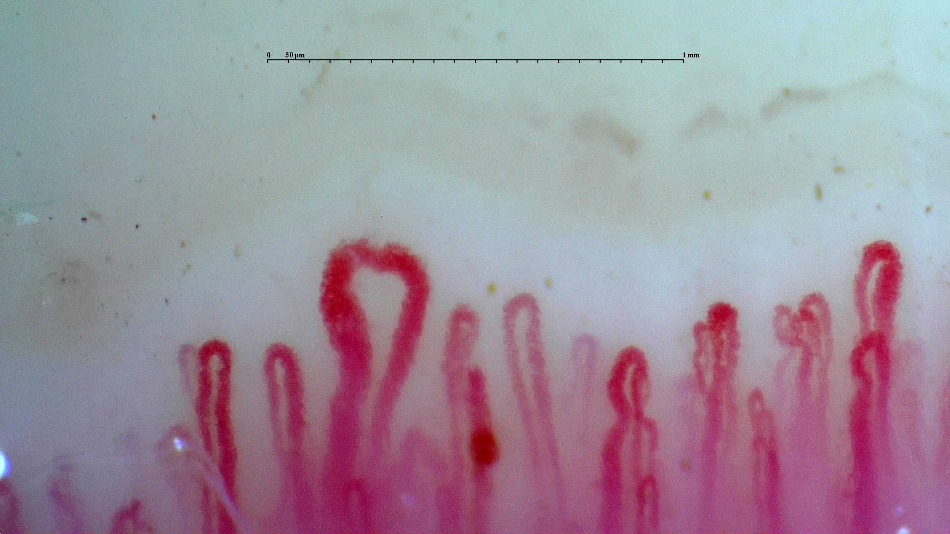
 Active scleroderma pattern: giant loop
A prominent enlarged loop in the active-pattern reference study. Compare the neighboring capillaries and the other fields before assigning a pattern.
View the complete studyHow to assess and measure
Record giants and hemorrhages, quantify distal-row density over calibrated lengths, and assess organization across several fingers. Report heterogeneity when fields differ. The ACA-positive and anti-Scl-70-positive cases below contain study-level quantitative findings; those values should not be assigned to every illustrated field.
Interpretation and related diseases
In systemic sclerosis, an active pattern is a useful morphological description, but it does not by itself define current activity in every organ. Dermatomyositis and overlap disease may show related microvascular findings. The complete cases demonstrate how serology, clinical context and image interpretation fit together.
Read the detailed clinical chapterCommon mistakes
- Choosing the subtype from the most dramatic loop alone.
- Equating an active capillaroscopic pattern with a universal measure of systemic disease activity.
- Requiring avascular areas: the anti-Scl-70 case reports an active pattern without them.
Real images and comparisons
Captions distinguish the illustrated finding from the study interpretation. Comparisons include variants, other clinical contexts or artifacts where available.
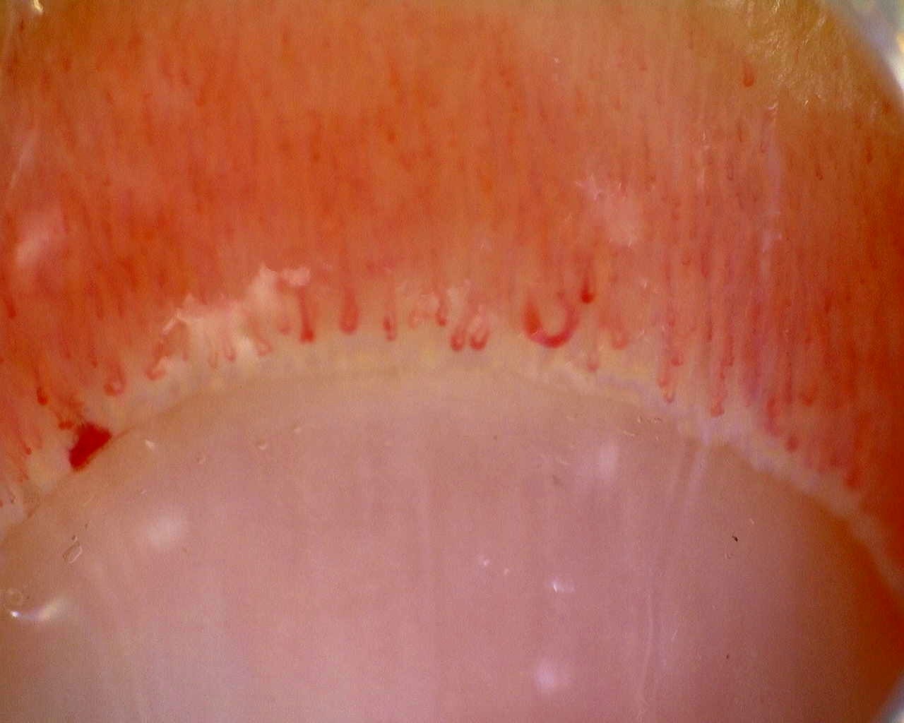 ACA-positive systemic sclerosis: overview
Overview of the nailfold in the ACA-positive active-pattern case. The reported density and giant-capillary percentage describe the study, not this photograph alone.
View the complete study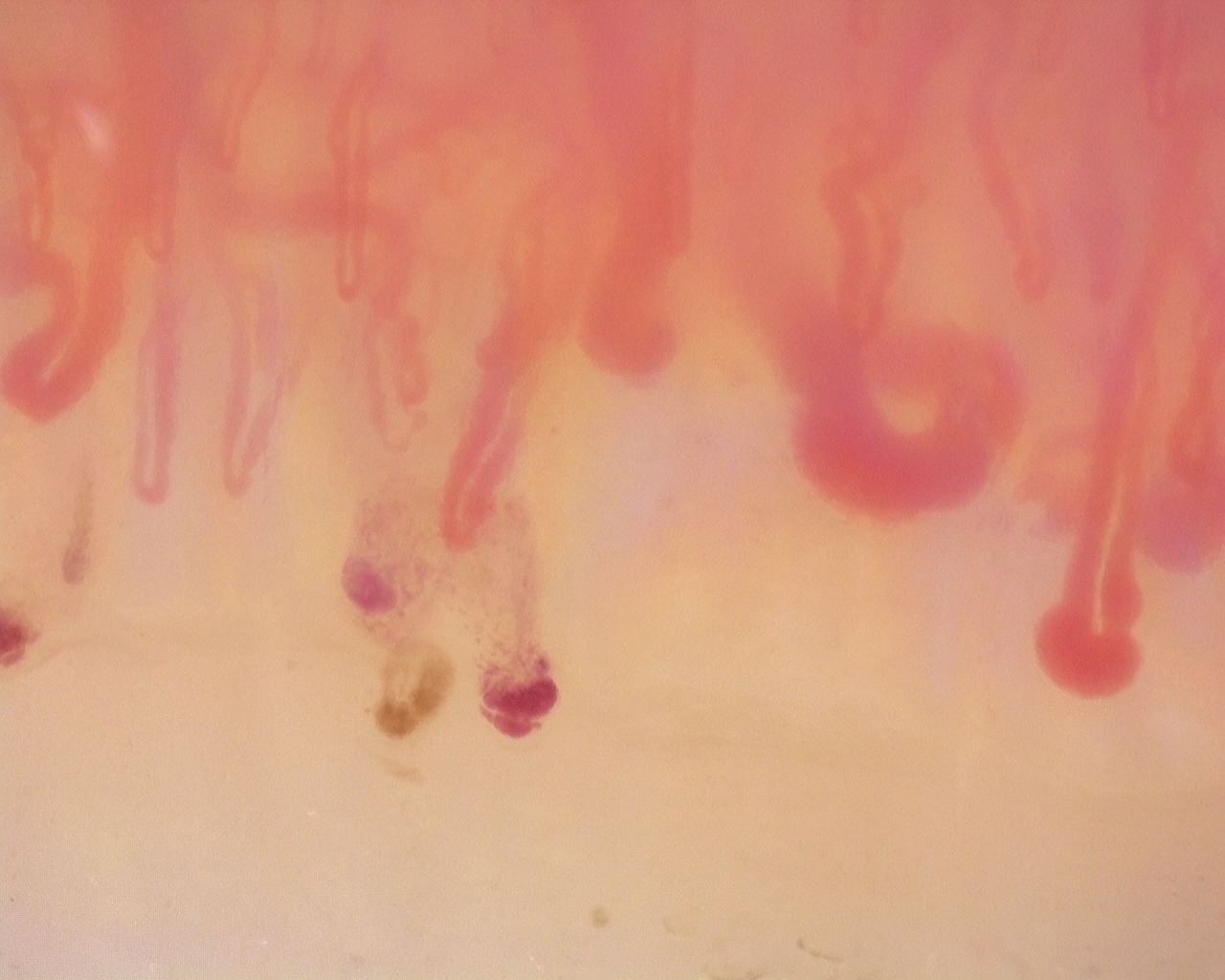 Anti-Scl-70 systemic sclerosis: enlarged loops and hemorrhages
Enlarged loops and dark hemorrhagic deposits in the anti-Scl-70-positive active-pattern case. The source report notes no avascular areas in the study.
View the complete study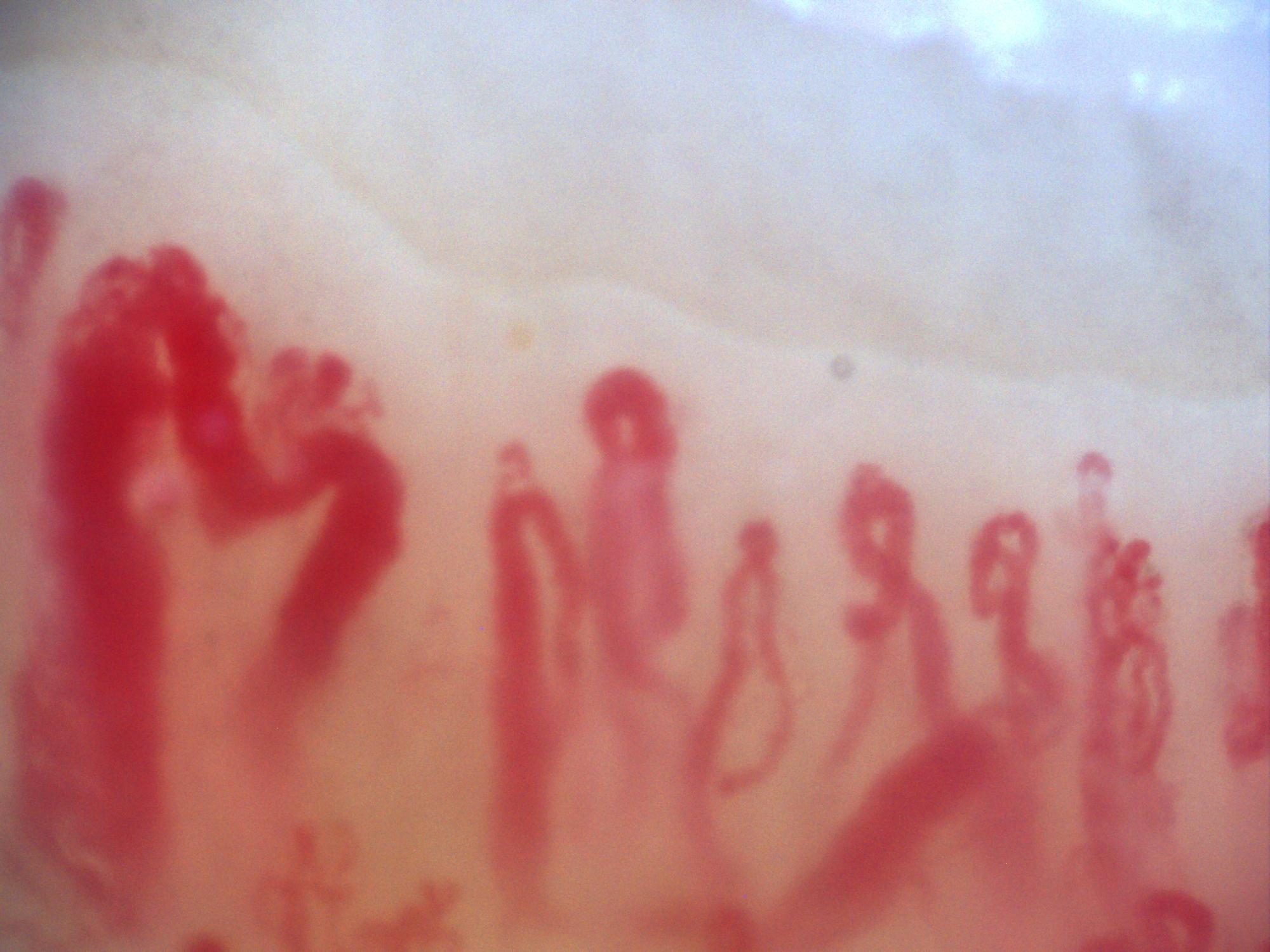
200x capillaroscopy image shared by Miguel Antonio Mesa Navas as an example of an active background with prominent megacapillaries.
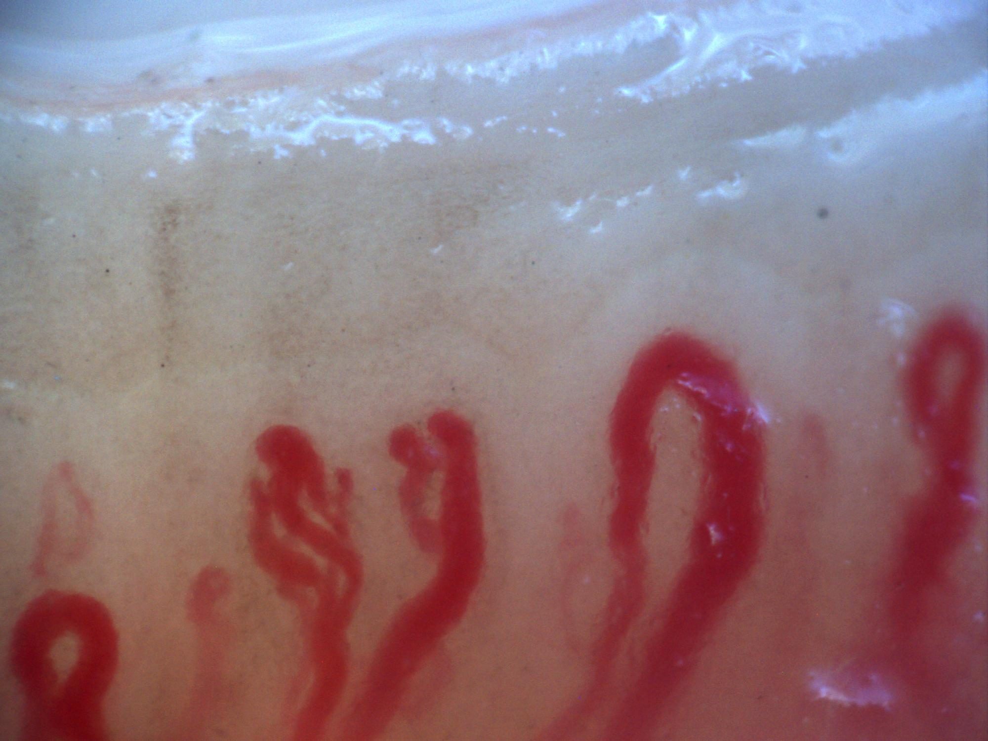
Another 200x capillaroscopy image shared by Miguel Antonio Mesa Navas as an example of an active background with prominent megacapillaries.
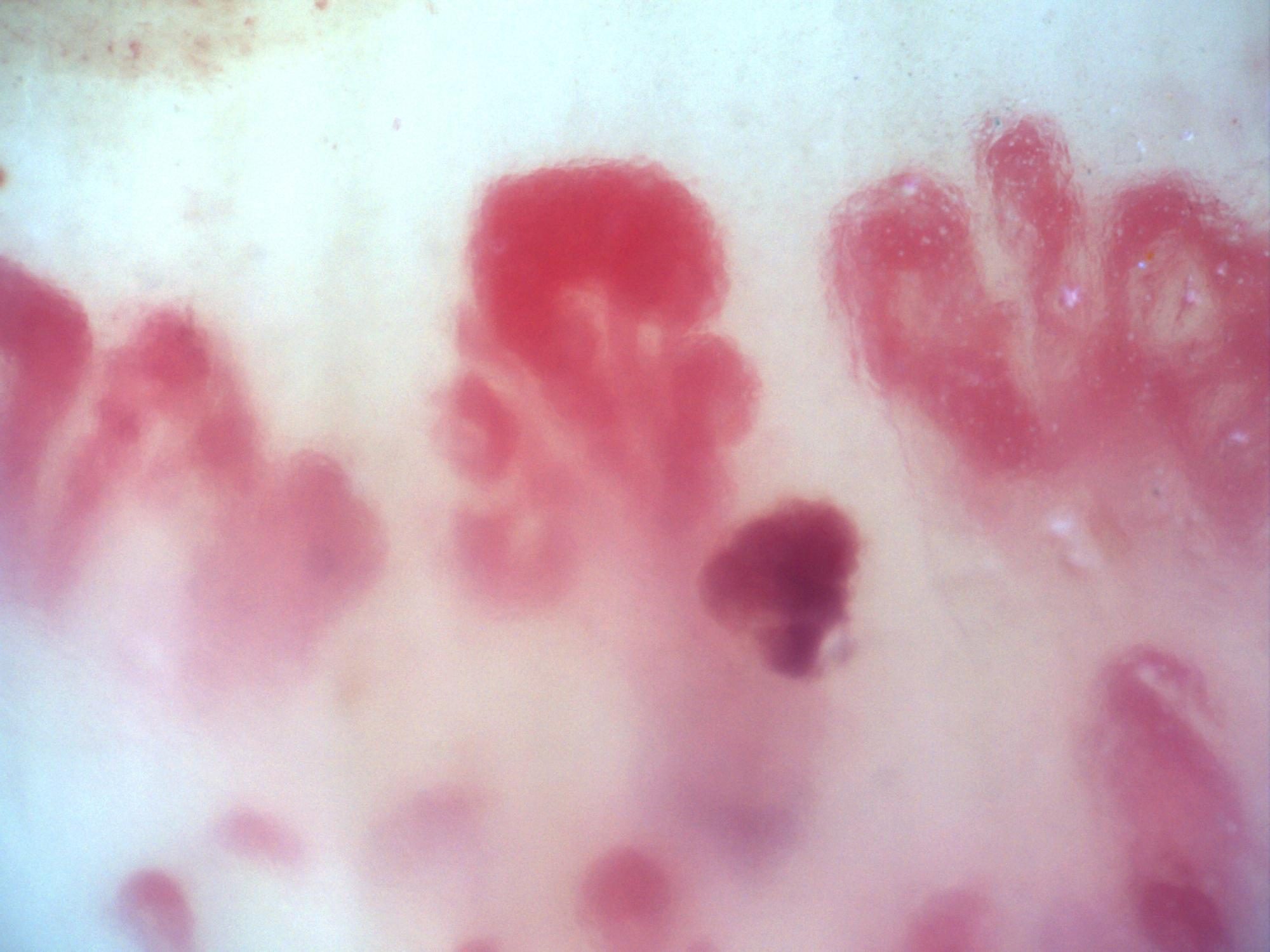
200x capillaroscopy image shared as an additional example of megacapillaries on an active background.
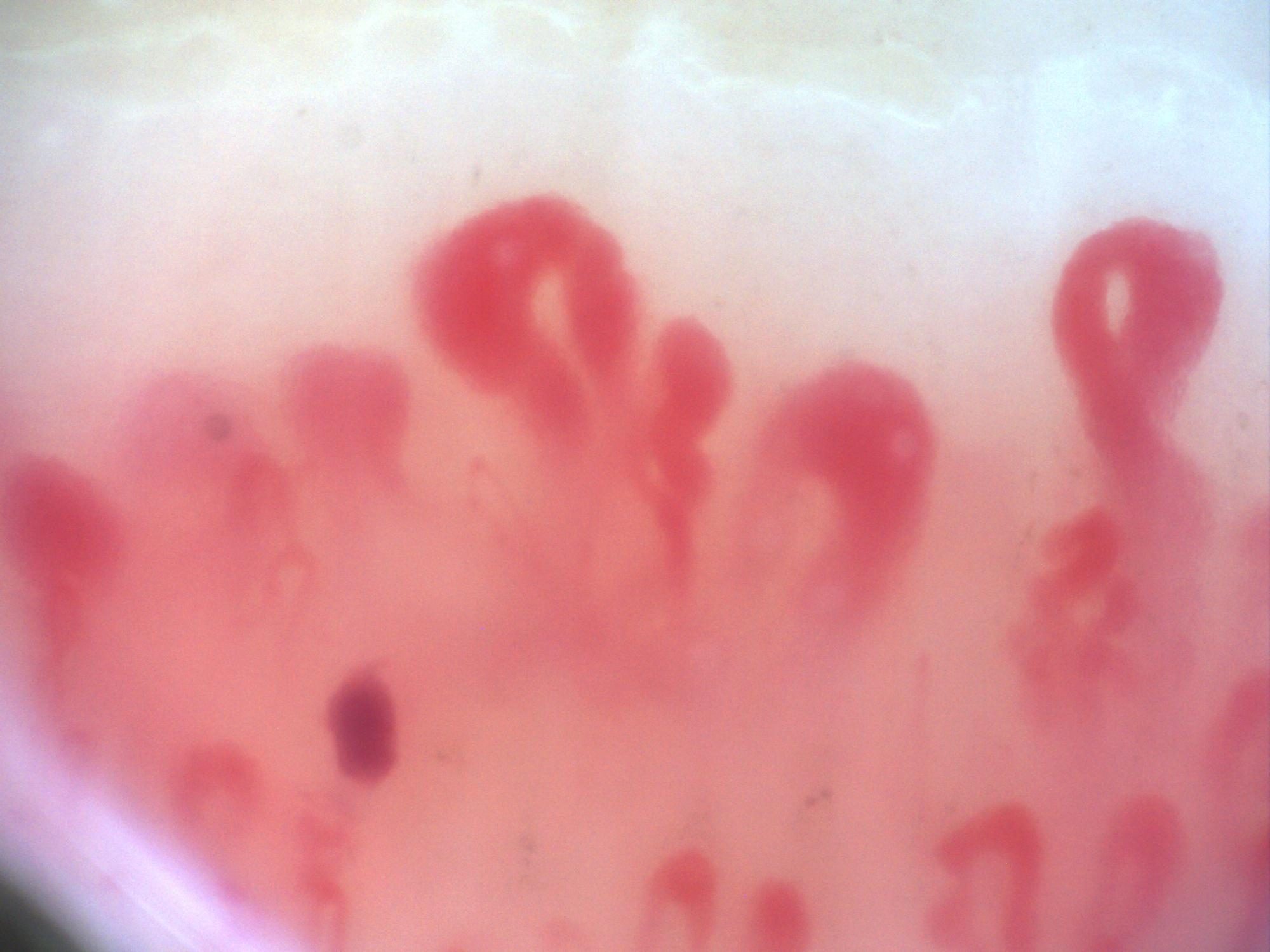
200x capillaroscopy image sent by Miguel Antonio Mesa Navas within the series of megacapillary examples.
Complete cases and studies
Active SSc Cutolo Pattern: Representative Case
The study shows frequent giant capillaries and hemorrhages with reduced density and early architectural disorganization.
Limited Cutaneous Systemic Sclerosis (ACA+) with Active Cutolo Pattern
Capillaroscopy shows marked capillary loss (5.3/mm), frequent dilations, high prevalence of giant capillaries (27.5%), and hemorrhages, matching an active Cutolo pattern.
Diffuse Systemic Sclerosis (Anti-Scl-70+) with Active Cutolo Pattern
Capillaroscopy shows marked capillary reduction (5.86/mm), frequent dilations (54%), a high prevalence of giant capillaries (39.7%), and hemorrhages, without avascular areas, matching an active Cutolo pattern. The CSURI index has increased. Blood flow is homogeneous, with reduced linear velocity.
Anticentromere-Positive Systemic Sclerosis with Inter-Digit Pattern Heterogeneity
The study is notable for pronounced inter-digit and inter-field heterogeneity. L4C shows marked capillary loss (desertification), whereas L4A has relatively preserved, normal-appearing architecture. R2C and L3D show giant capillaries within a disorganized architecture. R4A demonstrates late-stage revascularization or neoangiogenesis—a morphology the contributor describes as a "post-late" pattern—while R5A shows transitional active-to-late features. The complete study is classified as a late scleroderma pattern, with a mean capillary density of 5.63/mm, enlarged capillaries in 43.5%, and giant capillaries in 2.5%. Image quality is variable; several fields are technically limited and should not be interpreted in isolation.
References
- Smith V, et al. Standardisation of nailfold capillaroscopy for the assessment of patients with Raynaud’s phenomenon and systemic sclerosis. Autoimmun Rev. 2020;19:102458.
- Cutolo M, et al. Nailfold videocapillaroscopy assessment of microvascular damage in systemic sclerosis. J Rheumatol. 2000;27:155–160.
- Ingegnoli F, et al. Reporting items for capillaroscopy in clinical research on musculoskeletal diseases: a systematic review and international Delphi consensus. Rheumatology. 2021;60:1410–1418.
Scientific advisory board and contributors · Editorial policy