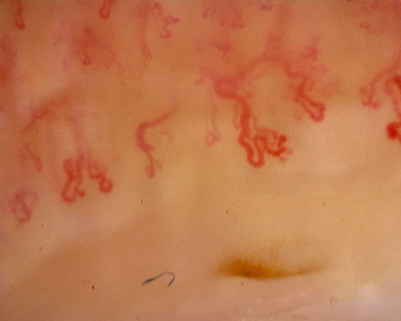
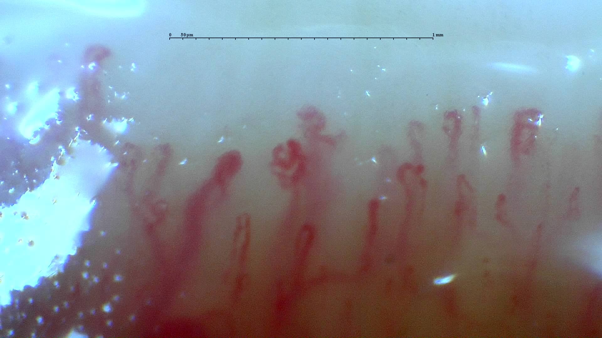
Anti-TIF1γ dermatomyositis: complex vascular shapes
Complex, winding and ramified vascular forms in the anti-TIF1γ dermatomyositis case. A scleroderma-like image appearance does not establish systemic sclerosis.
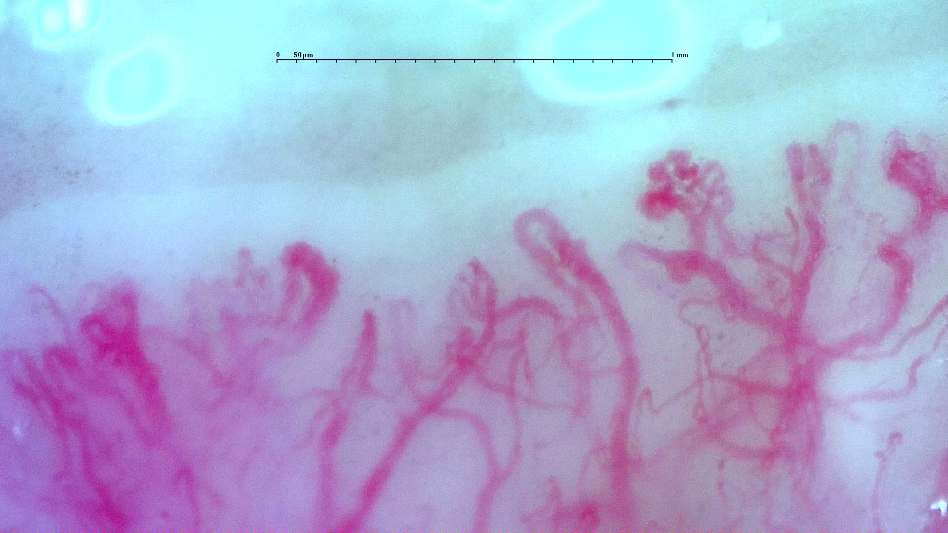
Source study context
Capillaroscopy shows severely reduced capillary density (2.7/mm), architectural disorganization, frequent dilations, giant capillaries, and abnormal/ramified forms (2/3 on the semi-quantitative scale), consistent with a scleroderma-like pattern.
Measurements and classification refer to the described study or visit; they are not new measurements of this image.
View the complete case and studyImage details
- Pathology
- Dermatomyositis (anti-TIF1γ+)
- Pattern
- Scleroderma-Like Pattern
- Severity
- See complete study
- Author or contributor
- Franklin Uguña Sari
- Hallmarks
- Abnormal shapes and ramifications
Teaching point
Complex, winding and ramified vascular forms in the anti-TIF1γ dermatomyositis case. A scleroderma-like image appearance does not establish systemic sclerosis.
Clinical caveat
This image illustrates a visual finding and should be interpreted with acquisition quality, distribution across fingers, clinical context, and serology when relevant.
Use and licensing
Educational material. For reuse outside Capillaroscopy.com, request permission and preserve the stated attribution.