Mixed connective tissue disease: initial overview
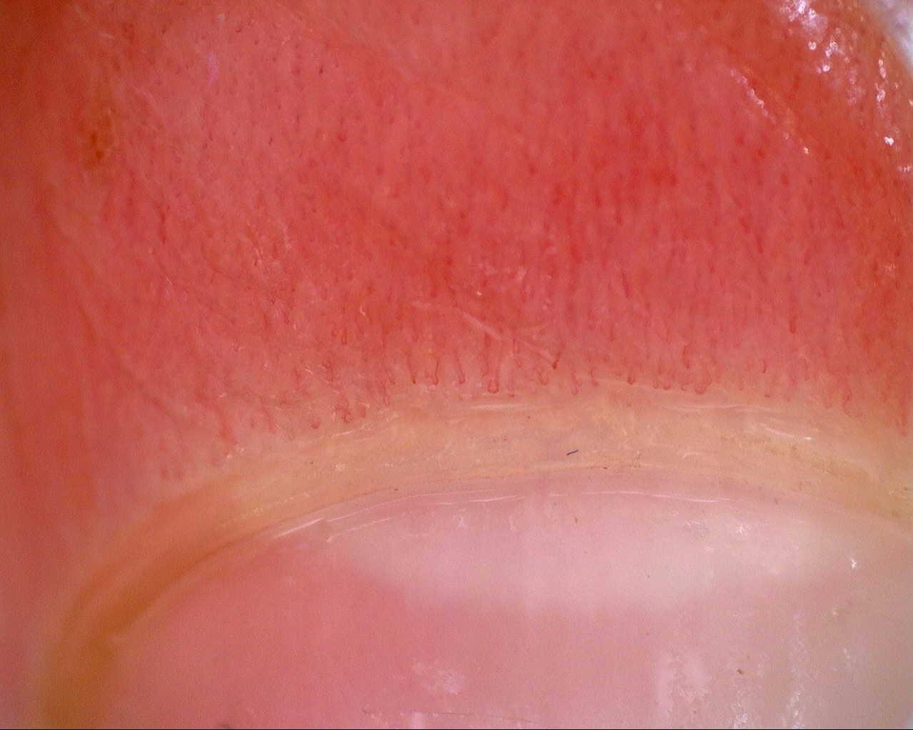
Initial nailfold overview from the longitudinal mixed connective tissue disease case. Use the serial studies to assess change in morphology and density.
Source study context
The initial capillaroscopy showed an early scleroderma pattern, supporting the diagnosis in the appropriate clinical and serological context. On follow-up, clinical improvement was accompanied by improvement of capillaroscopic findings, with higher density, disappearance of giant capillaries, and evolution toward a non-specific pattern.
Measurements and classification refer to the described study or visit; they are not new measurements of this image.
View the complete case and studyImage details
- Pathology
- Mixed Connective Tissue Disease (anti-RNP+)
- Pattern
- Early Scleroderma Pattern with Improvement
- Severity
- See complete study
- Author or contributor
- Clara Udaondo
Teaching point
Initial nailfold overview from the longitudinal mixed connective tissue disease case. Use the serial studies to assess change in morphology and density.
Clinical caveat
This image illustrates a visual finding and should be interpreted with acquisition quality, distribution across fingers, clinical context, and serology when relevant.
Use and licensing
Educational material. For reuse outside Capillaroscopy.com, request permission and preserve the stated attribution.
Related images
Back to atlasThere are not enough related images in this library yet.