Inter-digit heterogeneity: selected abnormal field
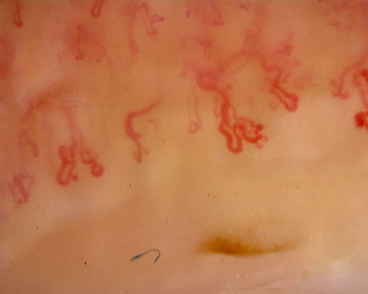
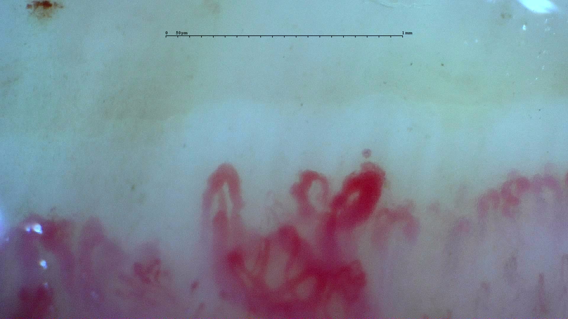
A disorganized field with enlarged loops and surface reflections from the inter-digit heterogeneity case. Other fields differ markedly; inspect the full study and its image-quality caveats.
Source study context
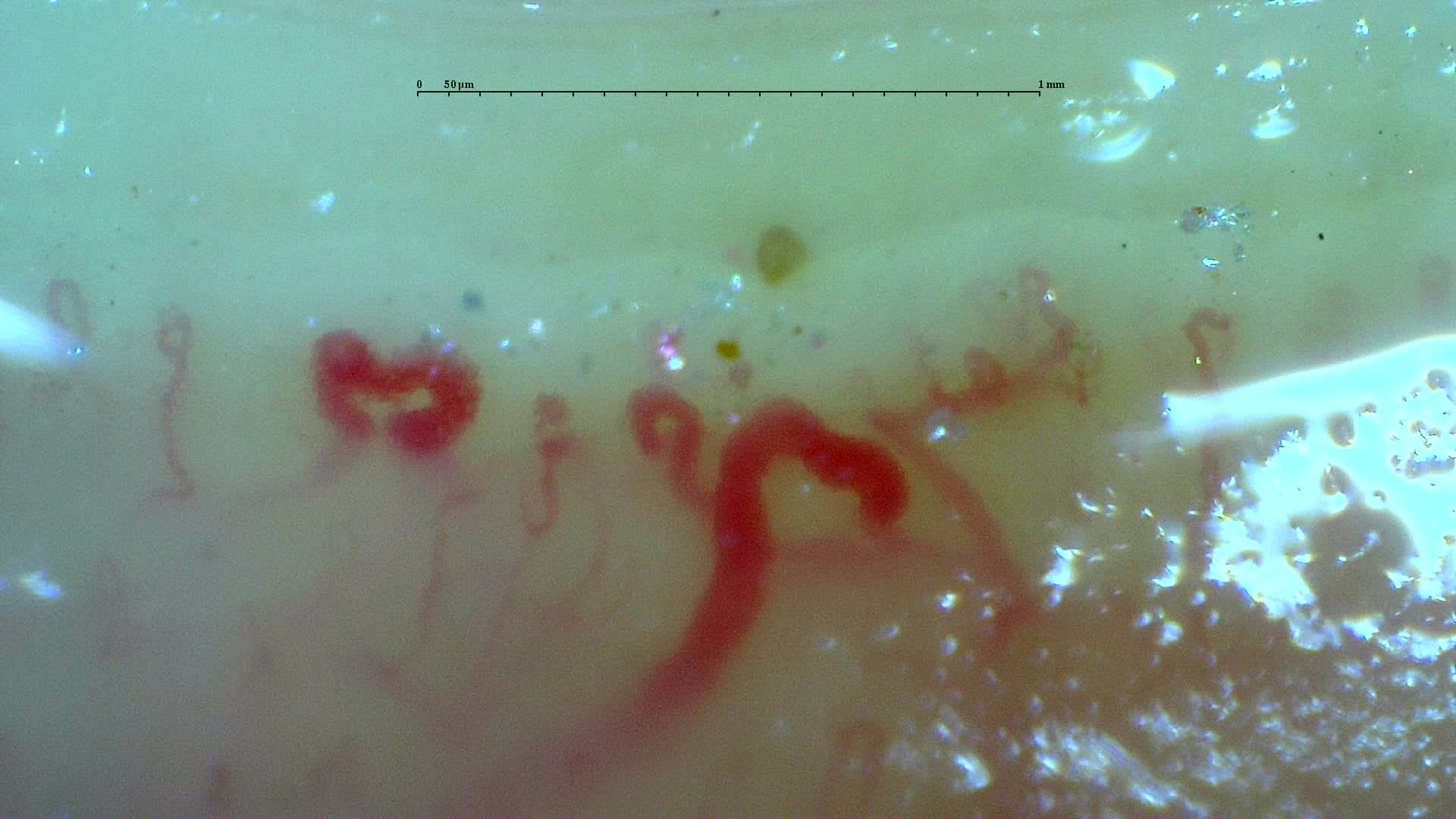
The study is notable for pronounced inter-digit and inter-field heterogeneity. L4C shows marked capillary loss (desertification), whereas L4A has relatively preserved, normal-appearing architecture. R2C and L3D show giant capillaries within a disorganized architecture. R4A demonstrates late-stage revascularization or neoangiogenesis—a morphology the contributor describes as a "post-late" pattern—while R5A shows transitional active-to-late features. The complete study is classified as a late scleroderma pattern, with a mean capillary density of 5.63/mm, enlarged capillaries in 43.5%, and giant capillaries in 2.5%. Image quality is variable; several fields are technically limited and should not be interpreted in isolation.
Measurements and classification refer to the described study or visit; they are not new measurements of this image.
View the complete case and studyImage details
- Pathology
- Systemic Sclerosis (Anticentromere-Positive)
- Pattern
- Heterogeneous Scleroderma Pattern (Active to Late)
- Severity
- See complete study
- Author or contributor
- Miguel Antonio Mesa Navas
- Hallmarks
- Late scleroderma pattern
Teaching point
A disorganized field with enlarged loops and surface reflections from the inter-digit heterogeneity case. Other fields differ markedly; inspect the full study and its image-quality caveats.
Clinical caveat
This image illustrates a visual finding and should be interpreted with acquisition quality, distribution across fingers, clinical context, and serology when relevant.
Use and licensing
Educational material. For reuse outside Capillaroscopy.com, request permission and preserve the stated attribution.