Image Library

Searchable collection of capillaroscopic images organized by pattern, diagnosis, and severity.

Selected examples

Start exploring

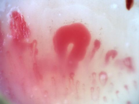
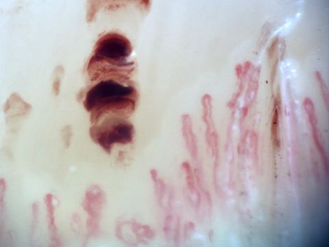
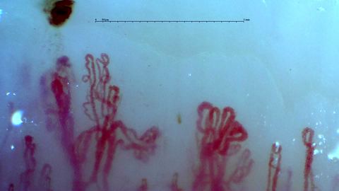
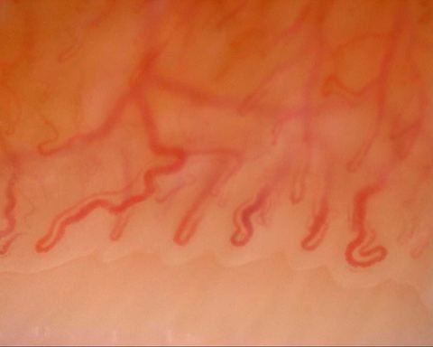
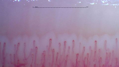
Compare capillary shapes, sizes and hemorrhages.

Browse all images (243) →
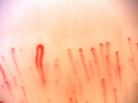
Broad, folded and branching-appearing capillaries with an elongated brown-red hemorrhage above them.
View details

A dark hemorrhagic deposit above complex loops

Author: Borja Gracia Tello

Magnification:
250×
Device:
Smart G-Scope
Field of view (W × H):
≈ 2.26 × 1.27 mm
Hemorrhages
Long hairpin capillaries with some enlarged rounded apices. Small brown deposits compatible with microhemorrhages lie near the lower left.
View details

Small brown foci beneath long parallel loops

Author: Borja Gracia Tello

Magnification:
200×
Device:
DinoLite
Hemorrhages
Hairpin capillaries of different widths with a small curved brown deposit compatible with a microhemorrhage above the center.
View details

A single small deposit above a varied capillary row

Author: Borja Gracia Tello

Magnification:
250×
Device:
Smart G-Scope
Field of view (W × H):
≈ 2.26 × 1.27 mm
Hemorrhages
Capillaries with rounded apices and tortuous limbs. An elongated hemorrhagic deposit and smaller brown foci lie above the row.
View details

Separate hemorrhagic foci along a sloping border

Author: Borja Gracia Tello

Magnification:
250×
Device:
Smart G-Scope
Hemorrhages
Mostly fine hairpin capillaries with two separate dark red microhemorrhages above the row.
View details

Two dark deposits above mostly fine loops

Author: Borja Gracia Tello

Magnification:
250×
Device:
Smart G-Scope
Field of view (W × H):
≈ 2.34 × 1.32 mm
Hemorrhages
Enlarged hairpin capillaries and some tortuous loops. Several dark hemorrhagic deposits form short rows above them.
View details

Rows of dark deposits above widened hairpins

Author: Borja Gracia Tello

Magnification:
250×
Device:
Smart G-Scope
Field of view (W × H):
≈ 2.39 × 1.34 mm
Hemorrhages
Mostly simple hairpin capillaries with one small coiled apex. An isolated elongated hemorrhage lies above the left side.
View details

An isolated dark streak above an open capillary row

Author: Borja Gracia Tello

Magnification:
250×
Device:
Smart G-Scope
Systemic lupus erythematosusHemorrhages
Fine elongated capillaries with numerous brown-red hemorrhagic deposits of different sizes.
View details

Multiple hemorrhagic deposits in an oblique row

Author: Borja Gracia Tello

Magnification:
207.55×
Device:
Dino-Lite Edge
Field of view (W × H):
≈ 1.88 × 1.50 mm
Hemorrhages
Capillaries with enlarged rounded apices and three separate reddish hemorrhagic deposits above the row.
View details

Three separated deposits above broad rounded loops

Author: Borja Gracia Tello

Magnification:
250×
Device:
Smart G-Scope
Field of view (W × H):
≈ 2.33 × 1.31 mm
Raynaud phenomenonHemorrhages
Elongated hairpin capillaries of different widths, with several small microhemorrhages above the left half.
View details

Small hemorrhagic foci over a closely spaced row

Author: Borja Gracia Tello

Magnification:
250×
Device:
Smart G-Scope
Systemic lupus erythematosusHemorrhages
Broad capillaries with elongated and rounded hemorrhagic deposits above the center and right side.
View details

Long hemorrhagic streaks and compact red deposits

Author: Borja Gracia Tello

Magnification:
250×
Device:
Smart G-Scope
Field of view (W × H):
≈ 2.29 × 1.29 mm
Hemorrhages
Slender capillary loops with a large dark red hemorrhagic deposit and smaller nearby foci at the upper right.
View details

A large dark hemorrhagic deposit with smaller nearby foci

Author: Borja Gracia Tello

Magnification:
200×
Device:
Optilia Digital Capillaroscope
Field of view (W × H):
≈ 1.71 × 1.28 mm
Hemorrhages
A long tortuous capillary on the right, beside straighter loops. Several small hemorrhagic deposits lie above the left half.
View details

Small hemorrhagic deposits beside a long tortuous loop

Author: Borja Gracia Tello

Magnification:
250×
Device:
Smart G-Scope
Field of view (W × H):
≈ 2.32 × 1.31 mm
Raynaud phenomenonHemorrhages
Capillaries with rounded apices of different widths and some irregular limbs. Brown-red hemorrhagic deposits form a vertical row near the center-right.
View details

A vertical row of deposits between capillary loops

Author: Borja Gracia Tello

Magnification:
200×
Device:
Optilia
Field of view (W × H):
≈ 1.71 × 1.28 mm
Hemorrhages
Rounded capillary loops with some curved limbs and a dense dark hemorrhagic deposit above the right side.
View details

A dark marginal deposit above rounded loops

Author: Borja Gracia Tello

Magnification:
200×
Device:
Optilia Digital Capillaroscope
Field of view (W × H):
≈ 1.71 × 1.28 mm
Hemorrhages
Fine and markedly enlarged capillaries, some partly obscured. An elongated dark hemorrhagic deposit lies above the left side.
View details

A dark deposit above loops with marked caliber variation

Author: Borja Gracia Tello

Magnification:
250×
Device:
Smart G-Scope
Field of view (W × H):
≈ 2.24 × 1.26 mm
Hemorrhages
Mostly simple hairpin capillaries with rounded apices. Several dark hemorrhagic deposits lie above the center-right.
View details

Vertically arranged hemorrhagic deposits above simple loops

Author: Borja Gracia Tello

Magnification:
250×
Device:
Smart G-Scope
Field of view (W × H):
≈ 2.35 × 1.32 mm
Hemorrhages
Fine hairpin capillaries with mild tortuosity and an isolated brown deposit compatible with a microhemorrhage above the row.
View details

An isolated curved brown deposit above tortuous loops

Author: Borja Gracia Tello

Magnification:
250×
Device:
Smart G-Scope
Hemorrhages
Capillaries with enlarged rounded apices and some folded loops. Several dark and pale red hemorrhagic deposits lie above them.
View details

Scattered hemorrhages above broad and twisted loops

Author: Borja Gracia Tello

Magnification:
250×
Device:
Smart G-Scope
Field of view (W × H):
≈ 2.37 × 1.33 mm
Antiphospholipid syndromeSystemic lupus erythematosusHemorrhages
Tortuous capillaries mixed with finer rounded hairpin loops. A thin brown hemorrhagic deposit lies above the center.
View details

A thin brown streak above tortuous capillaries

Author: Borja Gracia Tello

Magnification:
250×
Device:
Smart G-Scope
Field of view (W × H):
≈ 2.22 × 1.25 mm
Hemorrhages
Fine hairpin capillaries with multiple dark hemorrhagic deposits, including several elongated deposits near the center-right.
View details

Dense hemorrhagic streaks above fine loops

Author: Borja Gracia Tello

Magnification:
250×
Device:
Smart G-Scope
Hemorrhages
Enlarged hairpin capillaries mixed with finer loops. Irregular brown hemorrhagic deposits lie above the right side.
View details

Brown hemorrhagic patches above broadened tips

Author: Borja Gracia Tello

Magnification:
250×
Device:
Smart G-Scope
Field of view (W × H):
≈ 2.24 × 1.26 mm
Hemorrhages
Short loops with occasional coiled apices and a visible deeper plexus. A small brown deposit above the right side may represent a microhemorrhage.
View details

A small deposit above a broad view of the vascular bed

Author: Borja Gracia Tello

Magnification:
250×
Device:
Smart G-Scope
Field of view (W × H):
≈ 2.36 × 1.33 mm
Hemorrhages
Long enlarged hairpin capillaries with a wider central apex. An irregular dark hemorrhagic deposit lies above the left side.
View details

An irregular hemorrhage above long broad hairpins

Author: Borja Gracia Tello

Magnification:
250×
Device:
Smart G-Scope
Hemorrhages

Image Usage Guidelines

The atlas is an educational resource. Reuse rights are specific to each image: consult its detail page and the Image licensing and reuse page linked in the footer.

Images explicitly marked CC BY 4.0 may be reused, adapted and published, including commercially, under that licence without additional permission. Other images retain their individual terms.

Consent for educational publication does not by itself establish consent for broader reuse. A copyright licence does not settle patient privacy or other applicable rights.