Ongoing apical hemorrhage in systemic lupus erythematosus
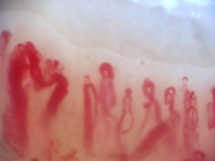
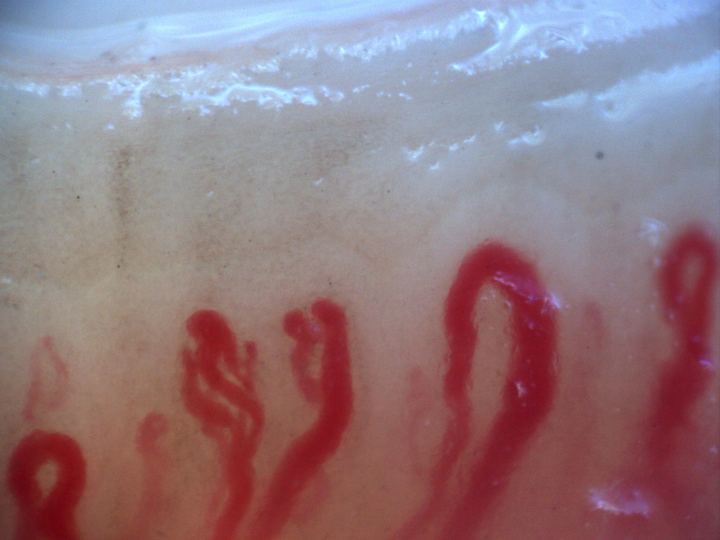
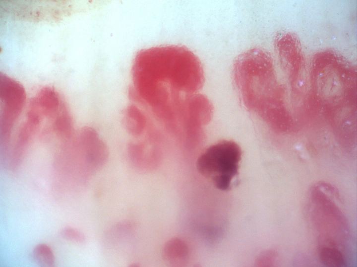
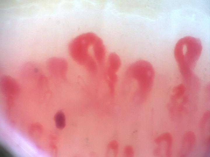
Nailfold capillaroscopy in systemic lupus erythematosus shows a scleroderma-like pattern. In the central field, a hemorrhage is forming around a capillary more than 50 μm in diameter. The pericapillary blood merges into a plume at the apex.
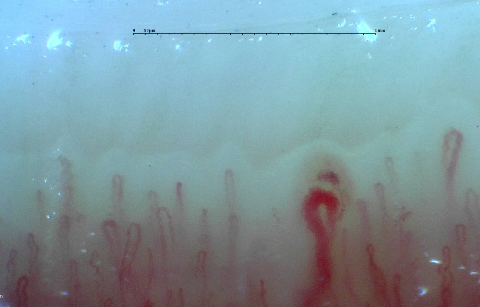
Image details
- Pathology
- Systemic lupus erythematosus
- Pattern
- Scleroderma-like pattern
- Author or contributor
- Miguel Antonio Mesa Navas
- Hallmarks
- Giant capillaryHemorrhages
- Magnification
- 250×
- Device
- Smart G-Scope
Teaching point
Follow the outline of the giant capillary to its apex, where the surrounding blood forms a plume.
Clinical caveat
This image illustrates a visual finding and should be interpreted with acquisition quality, distribution across fingers, clinical context, and serology when relevant.
Use and licensing
Contributed by Miguel Antonio Mesa Navas for educational publication on Capillaroscopy.com. For reuse permissions, contact the rights holder.
- Creator
- Miguel Antonio Mesa Navas