Giant-capillary morphology, hemorrhages and branching
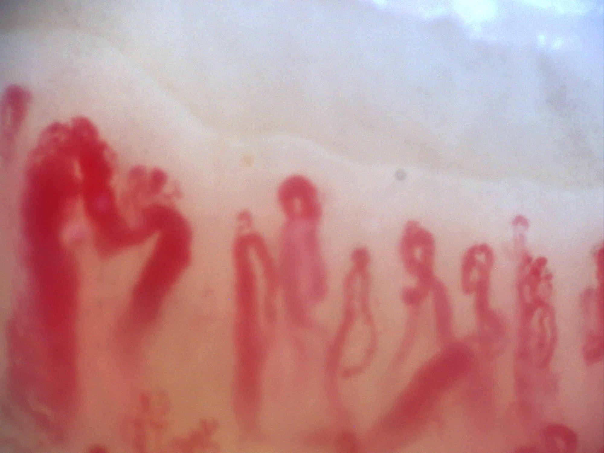
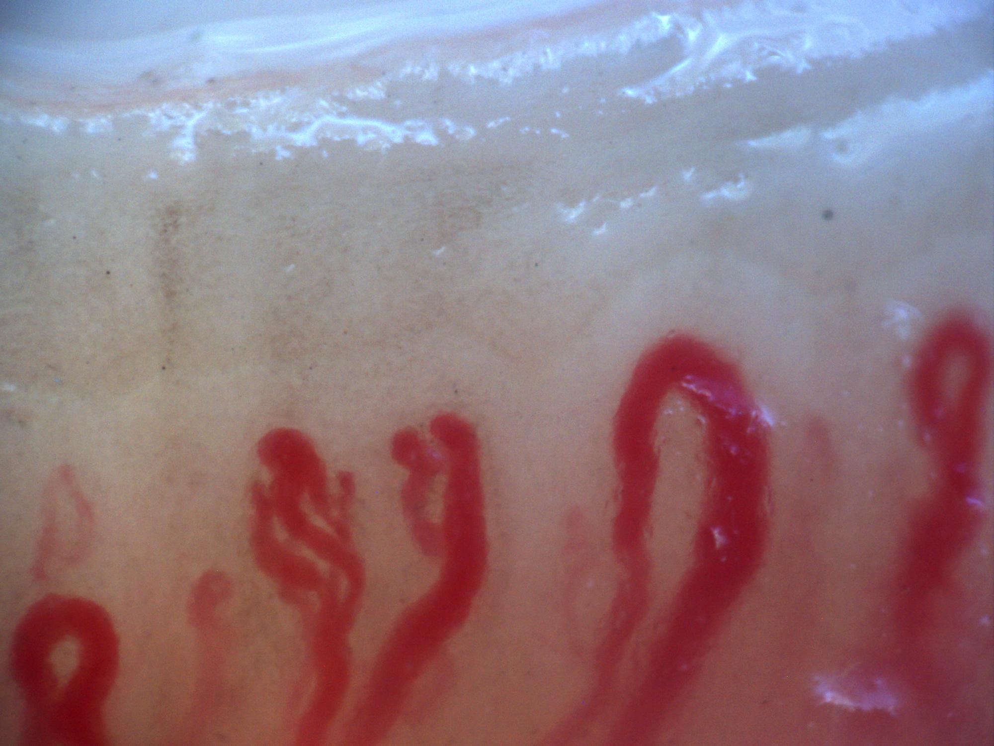
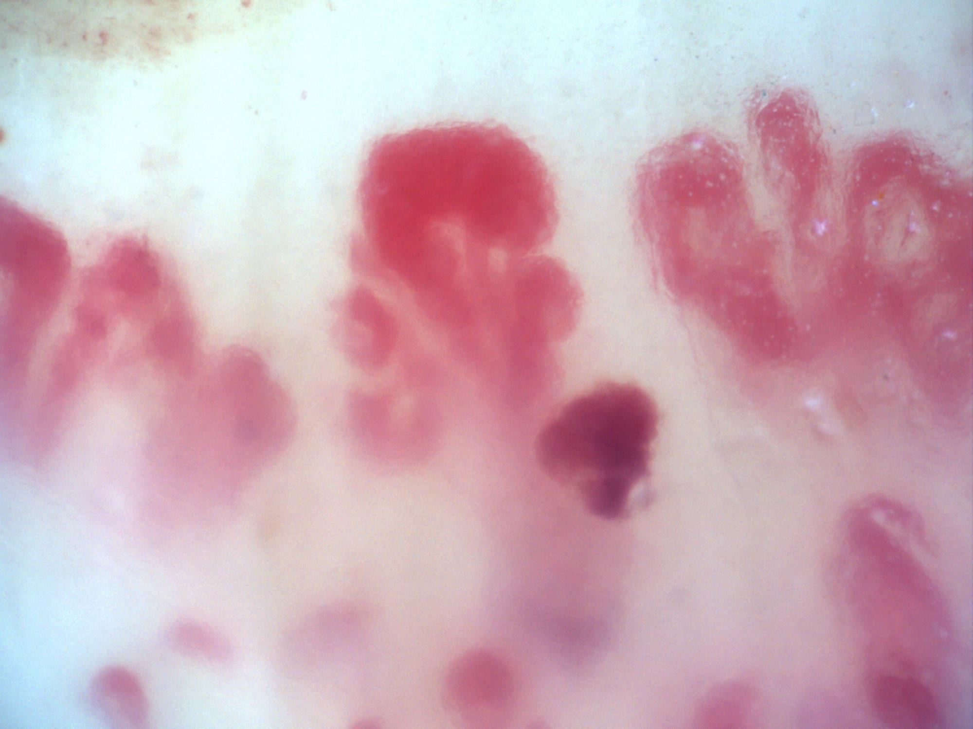
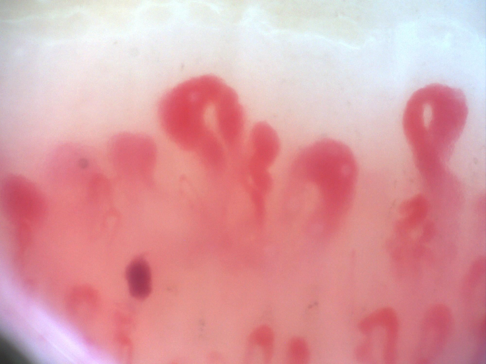
A giant-appearing oval capillary on the left and a small branching capillary on the right. Several hemorrhagic deposits lie near the center.
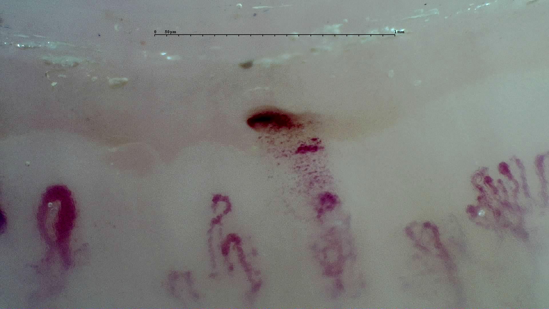
Image details
- Recorded diagnosis
- Limited systemic sclerosis
- Pattern
- Giant capillary
- Author or contributor
- Borja Gracia Tello
- Hallmarks
- Giant capillaryHemorrhagesAbnormal shapes and ramifications
- Magnification
- 250×
- Device
- Smart G-Scope
- Field of view (W × H)
- ≈ 2.30 × 1.29 mm
Teaching point
Describe whether abnormalities are isolated, repeated, or combined.
Measurements and calibration
For nonbranching apices with stable automated measurements, estimated widths span approximately 28–70 µm. These are automated estimates, not manually validated measurements.
Clinical caveat
This image illustrates a visual finding and should be interpreted with acquisition quality, distribution across fingers, clinical context, and serology when relevant.
Use and licensing
Educational material. For reuse outside Capillaroscopy.com, request permission and preserve the stated attribution.