Other vascular acrosyndromes: acrocyanosis, erythromelalgia, and differential diagnosis
An important part of clinical work is not calling non-Raynaud conditions Raynaud. Acrocyanosis, erythromelalgia, perniosis, paroxysmal digital hematoma, and digital necrosis may share acral color change, pain, or a relationship with temperature, but they arise from different mechanisms and require different decisions [1, 2].
- Raynaud is episodic and vasospastic; acrocyanosis is usually more persistent, and erythromelalgia is typically red, hot, and burning [1, 2, 5, 6, 7].
- Primary acrocyanosis is usually benign, but in a recent cohort, 45.3% of referred cases were secondary, and connective tissue diseases were the most common link [3].
- Capillaroscopy may be useful in acrocyanosis when it coexists with Raynaud, especially to avoid confusing its pattern with a scleroderma pattern [4].
- In erythromelalgia, capillaroscopy usually adds little; diagnosis remains clinical and treatment is stepwise, focused on triggers and the underlying cause [5, 6, 7].
- Perniosis, paroxysmal digital hematoma, and livedo are better recognized by their natural history and lesion morphology than by capillaroscopy [1, 2, 8, 9, 10].
- Digital necrosis, sustained pain, or a fixed lesion should not be managed as a trivial functional acrosyndrome: structural ischemia or vasculitis must be ruled out [1, 2].
Learning path
If you are unsure whether a case is Raynaud or one of its frequent mimics, this unit gives you the keys to avoid confusing them or overdiagnosing. Reading it together with Raynaud's phenomenon will help refine the differential diagnosis.
Quick differential diagnosis map
In acral disease, color and temperature are not enough. The most useful diagnostic approach combines trigger, duration, pain, persistence between episodes, and risk of tissue injury.
| Entity | Typical color | Main trigger | Usual duration | Pain | Risk of tissue loss |
|---|---|---|---|---|---|
| Raynaud's phenomenon | Pallor, with or without cyanosis and rubor | Cold or stress | Minutes to tens of minutes | Variable; more intense in secondary forms [1, 2] | Low in primary Raynaud, high if structural microangiopathy exists |
| Acrocyanosis | Persistent bluish color | Cold, vascular dependency | More persistent than episodic [1, 2, 3, 4] | Limited in primary forms; more relevant in secondary forms | Low in primary forms; higher if there is a secondary cause [3] |
| Erythromelalgia | Intense red and hot | Heat, exercise, limb dependency [5, 6, 7] | Minutes to hours | Prominent burning pain [5, 6, 7] | Low in most cases, but it can be highly disabling |
| Perniosis | Inflammatory erythematous-violaceous color | Damp cold | Days to weeks [1, 8] | Itching or pain | Usually low; look for secondary causes if persistent |
| Paroxysmal digital hematoma | Focal bluish-purple | Spontaneous or trivial trauma | Hours to a few days [9, 10] | Brief pain or paresthesia | Benign and self-limited |
| Digital necrosis | Fixed cyanosis, blackish discoloration, or ulceration | Vascular occlusion, vasculitis, critical ischemia | Persistent | Usually intense | High; diagnostic emergency [1, 2] |
The practical lesson is simple: the more persistent, fixed, unilateral, or destructive the presentation, the less likely it is to be a purely functional disorder. This rule of thumb helps distinguish benign from urgent situations even before ordering tests [1, 2].
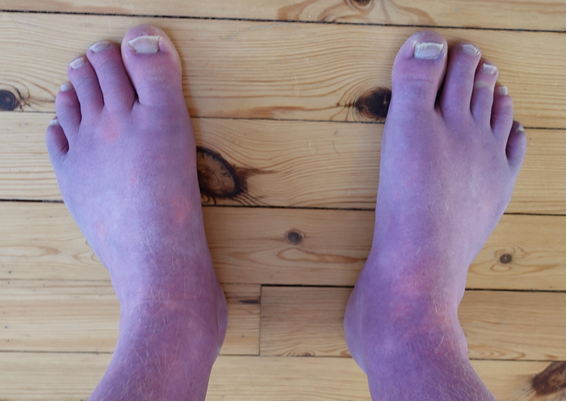
Acrocyanosis and acrorhigosis
Acrocyanosis is probably the most common "mimic" of Raynaud when the history is poor. Unlike episodic vasospasm, it is dominated by more persistent bluish discoloration, with cold and often sweaty hands and feet [1, 2, 3, 4].
Primary acrocyanosis is usually symmetric, chronic, relatively painless, and rarely causes necrosis. The problem is that not all acrocyanosis is primary. In a 2025 observational study, 24 of 53 patients referred for acrocyanosis, or 45.3%, had a secondary form. Age 40 years or older was associated with a relative risk (RR) of 2.5, and connective tissue diseases accounted for 71% of secondary forms [3]. In other words, although the entity is generally benign, it should not be trivialized if the clinical context is suspicious.
The term acrorhigosis is used less uniformly in contemporary literature. It usually refers to acral coldness with stiffness and marked vascular hypertonia, somewhere between functional vasospasm and persistent hypoperfusion. Current evidence is limited and terminology is not fully standardized, so it is preferable to describe the clinical phenotype rather than overuse a label that is not universal.
What capillaroscopy may show in acrocyanosis
Capillaroscopy is not the central test for diagnosing acrocyanosis, and it can be very useful when the patient also has Raynaud and the main question is avoiding overdiagnosis of a scleroderma pattern [1, 4]. In the 2024 French series focused on patients with acrocyanosis associated with Raynaud, capillary density remained normal in all but one patient, there were no avascular areas, and hemorrhages were very frequent, up to 82% of cases. Dilations were also observed, especially in the efferent venous limb, but only one isolated giant loop [4]. This combination does not fit the rarefaction typical of a scleroderma pattern.
Practically speaking: acrocyanosis can make the capillaroscopy look noisy or abnormal, but usually without important capillary loss or avascular areas. If the bed shows preserved density, venous dilation, and hemorrhages in a clinical context of persistent cyanosis and cold extremities, the overall picture does not by itself suggest a scleroderma pattern [1, 4].
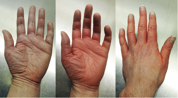
Erythromelalgia
In clinical terms, erythromelalgia is almost the thermal counterpart of Raynaud: episodes of erythema, heat, and burning pain, precipitated by heat or exercise and relieved by cooling [5, 6, 7].
A reference 2024 review describes it as a rare, underdiagnosed, and potentially highly disabling disorder [5, 6]. It may be idiopathic, hereditary, or secondary to systemic diseases, including myeloproliferative neoplasms, neuropathies, and some autoimmune contexts [5, 6, 7]. Unlike Raynaud, the patient often seeks cold rather than avoiding it.
Management focuses on confirming the diagnosis, acting on triggers, and looking for treatable secondary causes. Another important 2023 review emphasizes that universal guidelines do not exist and therapeutic response is highly variable; treatment is usually stepwise and combines behavioral measures with topical or systemic drugs selected according to the pain phenotype and underlying cause [7].
Patients should be warned about a practical risk: excessive cooling. Prolonged immersion in ice water or repeated ice use may provide temporary relief, but can also cause maceration, ulceration, or secondary skin injury [5, 6, 7], which is especially relevant in severe cases.
What is the role of capillaroscopy?
In erythromelalgia, capillaroscopy is not as decisive as it is in Raynaud. It may be normal or show nonspecific changes, so it rarely leads to the diagnosis by itself [1, 5, 6, 7]. However, the value of the test increases if the patient also reports vasospastic episodes compatible with Raynaud or if associated connective tissue disease is suspected.
Perniosis, livedo, and digital hematoma
These disorders are often confused when the clinician focuses only on color. What distinguishes them from Raynaud is lesion morphology and natural history.
Perniosis
Perniosis or chilblain is an acral inflammatory lesion induced by cold and humidity, not a brief vasospasm. Dubey's review emphasizes that lesions usually appear 12 to 24 hours after the trigger and persist for days or weeks, with painful or pruritic papules or plaques, sometimes erosive [8]. If the presentation is persistent, appears in warm seasons, leaves scars, or is accompanied by systemic findings, secondary forms should be sought, especially cutaneous or systemic lupus [1, 8].
Livedo reticularis
Livedo reticularis is recognized by a violaceous reticular pattern rather than by paroxysmal acral attacks. It may be physiologic, cold-induced, or secondary to vascular, hematologic, or autoimmune disease [1]. Capillaroscopy plays a modest role, and the priority is distinguishing benign forms from those with fixed retiform pattern, ulceration, or systemic findings.
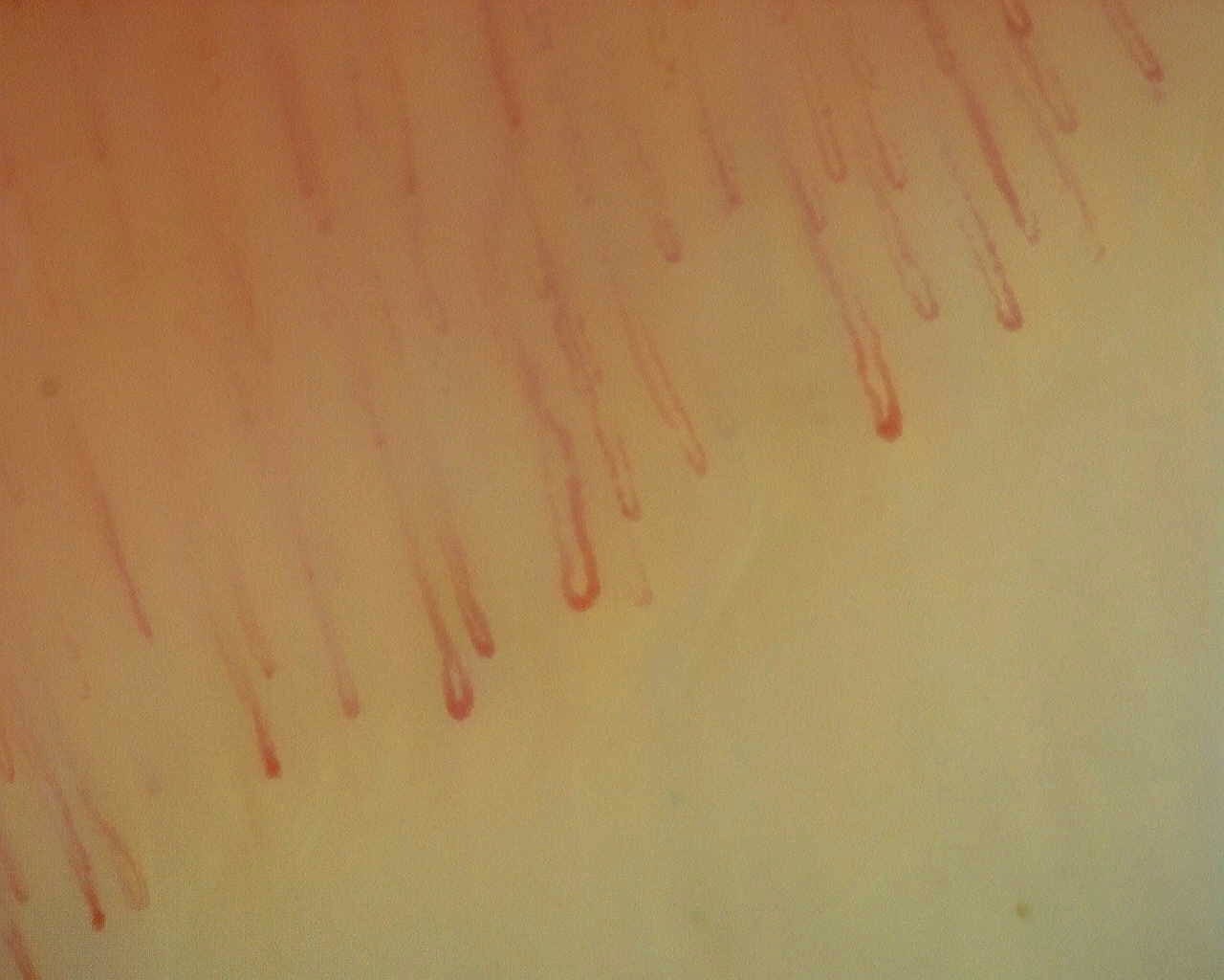
Paroxysmal digital hematoma or Achenbach syndrome
Achenbach syndrome is a benign but alarming cause of a "painful blue finger." It is characterized by sudden ecchymosis, often on the volar aspect of one or more fingers, with pain or paresthesias and spontaneous resolution within a few days [9, 10]. In most cases the fingertip is spared and vascular studies are normal, which helps separate it from acute arterial ischemia [9].
This diagnosis matters because it avoids unnecessary invasive tests. If the presentation is typical and there are no signs of vasculitis, embolism, or necrosis, management is conservative; reassuring the patient is important [9, 10].
Digital necrosis
Digital necrosis does not belong to the group of benign functional disorders. It requires consideration of arterial occlusion, vasculitis, antiphospholipid syndrome, cryoglobulinemia, thromboangiitis, complicated systemic sclerosis, or embolic damage, among others [1, 2]. Capillaroscopy can provide microvascular context, but it must not delay vascular and systemic evaluation, which should be undertaken promptly.

When capillaroscopy truly helps
Capillaroscopy does not perform equally across all acrosyndromes. Knowing when it adds value and when it does not can avoid low-yield requests.
| Entity | Capillaroscopy utility | Practical comment |
|---|---|---|
| Raynaud | High | Essential for separating primary forms from structural microangiopathy [1, 2] |
| Acrocyanosis | Moderate | Useful especially if it coexists with Raynaud and false positives for scleroderma pattern must be avoided [3, 4] |
| Erythromelalgia | Low | Normal or nonspecific in many cases; usually not the decisive test [1, 5, 6, 7] |
| Perniosis | Low to contextual | It is more useful to look for the clinical cause than to expect a defining capillary pattern [1, 8] |
| Achenbach | Very low | The typical clinical picture matters more than the microimage [9, 10] |
| Digital necrosis | Complementary | It must not delay urgent evaluation of the ischemic cause [1, 2] |
From a teaching perspective, this table summarizes a useful idea: capillaroscopy is excellent for Raynaud and much less decisive for several of its "mimics." Therefore, it should be requested with a concrete question, not simply as a reaction to any acral color change.
Short clinical cases
These examples show why the differential diagnosis is not merely an academic exercise, but an essential tool to avoid management errors.
Case 1: it was not Raynaud, it was acrocyanosis
A 19-year-old woman has cold, bluish hands almost all winter, without a clear pallor-rubor sequence. She reports hyperhidrosis and persistent color change rather than attacks. The history already points more to primary acrocyanosis than to Raynaud.
Case 2: acrocyanosis with clues to a secondary form
A 47-year-old patient has new-onset acrocyanosis, arthralgias, and positive antinuclear antibodies (ANA). Although acrocyanosis may be functional, this context better matches the high-yield profile for investigation of secondary disease described in recent series [3].
Case 3: benign blue finger
A 52-year-old woman develops sudden painful ecchymosis on the volar aspect of the fourth finger, with fingertip sparing and spontaneous resolution in 72 hours. The clinical picture is much more compatible with Achenbach syndrome than with Raynaud or digital embolism [9, 10].
Case 4: red, hot, and burning
A patient has burning pain in the feet that worsens with hot showers and improves by uncovering the feet. This is not a Raynaud pattern. The priority is to consider erythromelalgia and rule out secondary causes, including myeloproliferative neoplasms or neuropathy [5, 6, 7].
FAQ
Does acrocyanosis produce the same attacks as Raynaud?
No. Acrocyanosis is usually more persistent and less clearly paroxysmal, with cold and symmetric cyanosis [1, 2, 3].
Is acrocyanosis always benign?
Not always. In patients referred to specialized clinics, there may be a relevant proportion of secondary forms, especially with older age or systemic symptoms [3].
Can capillaroscopy look abnormal in acrocyanosis?
Yes, but the pattern usually preserves normal density and does not show avascular areas typical of systemic sclerosis [4].
Can erythromelalgia and Raynaud coexist?
Yes, although it is uncommon. Coexistence does not invalidate the diagnosis; it requires careful description of what triggers each type of episode and how it appears [5, 6, 7].
Is perniosis a type of Raynaud?
No. It is a cold-induced acral inflammatory lesion with a course of days or weeks and its own lesion morphology [1, 8].
Glossary
- Acrocyanosis
- Bluish acral discoloration, more persistent than episodic, generally symmetric and associated with local coldness.
- Erythromelalgia
- Paroxysmal disorder with erythema, heat, and burning pain, typically worsened by heat or exercise.
- Perniosis
- Cold- and damp-induced acral inflammatory lesions, also called chilblains.
- Achenbach syndrome
- Benign paroxysmal digital hematoma with painful ecchymosis and spontaneous resolution.
- Vascular acrosyndrome
- Umbrella term for acral disorders of color, temperature, or perfusion of functional or structural origin.
References
- Casanegra AI, Shepherd RF. Raynaud Phenomenon and Other Vasospastic Disorders. Cardiol Clin. 2021;39(4):583-599. doi: 10.1016/j.ccl.2021.06.010. PMID: 34686269.
- Zahn C, Puga C, Malik A, Khanna D. Painful Raynaud's mimics. Best Pract Res Clin Rheumatol. 2024;38(1):101948. doi: 10.1016/j.berh.2024.101948. PMID: 38704280.
- Bilancini S, Lucchi M, Trevisan G, Di Pino L, Tucci S. Acrocyanosis: primary or secondary form? An observational study. Acta Dermatovenerol Alp Pannonica Adriat. 2025;34(4):177-180. PMID: 41420618.
- Guelimi R, Monfort JB, Chaby G, Lok C, Lazareth I, Maillard H, et al. Nailfold capillaroscopy in acrocyanosis among patients with associated Raynaud's phenomenon. Ann Dermatol Venereol. 2024;151(3):103309. doi: 10.1016/j.annder.2024.103309.
- Gonzalez Caldito E, Kaul S, Gonzalez Caldito N, Piette W, Mehta S. Erythromelalgia. Part I: Pathogenesis, clinical features, evaluation, and complications. J Am Acad Dermatol. 2024;90(3):453-462. doi: 10.1016/j.jaad.2023.02.071. PMID: 37364617.
- Gonzalez Caldito E, Gonzalez Caldito N, Kaul S, Piette W, Mehta S. Erythromelalgia. Part II: Differential diagnoses and management. J Am Acad Dermatol. 2024;90(3):465-474. doi: 10.1016/j.jaad.2023.02.070. PMID: 37364616.
- Ma JE, Lee JUJ, Sartori-Valinotti JC, Rooke TW, Sandroni P, Davis MDP. Erythromelalgia: A Review of Medical Management Options and Our Approach to Management. Mayo Clin Proc. 2023;98(1):136-149. doi: 10.1016/j.mayocp.2022.08.005. PMID: 36470753.
- Dubey S, Joshi N, Stevenson O, Gordon C, Reynolds JA. Chilblains in immune-mediated inflammatory diseases: a review. Rheumatology (Oxford). 2022;61(12):4631-4642. doi: 10.1093/rheumatology/keac231. PMID: 35412601.
- Lehman H, Acho R, Hans SS. Achenbach syndrome as a rare cause of painful, blue finger. J Vasc Surg Cases Innov Tech. 2021;7(3):589-592. doi: 10.1016/j.jvscit.2021.06.016. PMID: 34541431.
- Yie K. Achenbach Syndrome: A Benign Painful Blue Finger with Tip Sparing. Vasc Specialist Int. 2019;35(4):251-253. doi: 10.5758/vsi.2019.35.4.251. PMID: 31915672.