In systemic lupus erythematosus (SLE), capillaroscopy rarely establishes the diagnosis. Its real value lies in the context of Raynaud's phenomenon, anti-U1-RNP, digital lesions, pulmonary arterial hypertension (PAH), and vascular activity, where microangiopathy may occur but is heterogeneous. Even the latest literature shows a mixture of normal examinations, nonspecific findings, and a minority of scleroderma patterns [1, 2, 3, 4, 5, 6, 7, 8, 9, 10, 11, 12].
- In SLE, capillaroscopy is a supporting technique, not a classification criterion or a substitute for clinical, immunological, or organ assessment [1, 2].
- The most consistent findings are modest: more hemorrhages, dilated or giant capillaries, and morphological abnormalities than in healthy controls, but most patients do not develop a specific reproducible pattern [2, 4, 12].
- In a recent international cohort assessed by blinded readers, 86.6% of patients with SLE retained an overall normal pattern, although abnormal findings were more frequent than in healthy controls [12].
- Raynaud's phenomenon, anti-U1-RNP, childhood-onset lupus, and suspected PAH are settings in which the test may add more than in stable SLE without vascular symptoms [3, 6, 7, 8, 9, 10, 11].
- A scleroderma pattern in SLE does not automatically mean overlap with systemic sclerosis; it calls for review of the complete phenotype, serology, and follow-up, but does not change the diagnosis on its own [1, 3, 8].
- Associations with activity, nephritis, or damage have been reported in some cohorts, but remain too variable to use capillaroscopy as an isolated biomarker for treatment monitoring [2, 4, 5, 6, 7, 11, 12].
Learning path
This unit explains what capillaroscopy can add in SLE without overstating its value: it is usually more useful for specific vascular problems than for classification. Then review APS and vasculitis to compare settings with even less specific findings [1, 2, 3, 8, 12].
What to expect in SLE
The most common error in SLE is looking for a capillaroscopic signature equivalent to the systemic sclerosis pattern. This expectation does not fit the available evidence and helps explain why findings are often overinterpreted [1, 2].
Cutolo et al's systematic review analyzed 40 studies and identified a methodological problem that persists: different techniques, different magnifications, inconsistent definitions, and small cohorts [2]. The underlying message was simple. SLE may show more hemorrhages, morphological abnormalities, and tortuous capillaries than healthy controls, but the direction and magnitude of quantitative findings vary across studies [2].
More recent studies do not eliminate this heterogeneity, but interpret it more clearly. An international cohort coordinated by the European Alliance of Associations for Rheumatology (EULAR) microcirculation group showed that most patients with SLE retain an overall normal pattern. Nevertheless, when abnormalities occur, dilated and giant capillaries, hemorrhages, and irregular architecture are more frequent than in healthy individuals [12]. These observations qualify the impressions from some older single-center studies, in which abnormality rates appeared very high [4].
Capillaroscopy in SLE is therefore more useful for contextualizing a specific vascular problem than for establishing the disease diagnosis. In a patient with arthritis, nephritis, or cytopenias but no Raynaud's phenomenon or vascular symptoms, the test is unlikely to change management. The balance shifts when Raynaud's phenomenon, anti-U1-RNP, digital lesions, or suspected PAH are present [1, 2, 3, 8, 9, 10, 11].
Recurring findings
Although there is no universal SLE pattern, some findings recur often enough to warrant standardized interpretation [2, 4, 11, 12].
The most frequently reported findings are hemorrhages, dilations, isolated giant capillaries, increased tortuosity, and architectural abnormalities. Among 85 newly diagnosed patients, Zhao et al found a normal pattern in 15.3%, a nonspecific pattern in 75.3%, and a scleroderma pattern in 9.4% [4]. At the other extreme, the 2025 international cohort reported a normal pattern in 86.6% overall. Prevalence therefore depends substantially on the methods and the threshold used to classify a test as abnormal [12].
This divergence does not invalidate the observations, but calls for interpretation by individual domains. A useful SLE capillaroscopy report should describe whether pathological hemorrhages, enlarged or giant capillaries, reproducibly reduced density, and architectural disorganization are present. Avoid forcing every combination of minor findings into a fixed diagnostic label [1, 2, 12].
| Domain | Reported findings | Practical interpretation |
|---|---|---|
| Hemorrhages | More frequent than in controls and sometimes associated with activity or nephritis [2, 4, 6, 7, 11, 12] | Relevant when repeated and well defined, rather than isolated or trauma-related |
| Dilated or giant capillaries | Present in a minority; more likely in phenotypes with Raynaud's phenomenon or high activity [4, 11, 12] | Do not imply systemic sclerosis on their own |
| Tortuosity and irregular architecture | Very frequent, but poorly specific [2, 4, 12] | Describe without overstating |
| Capillary density | Conflicting results across studies [2, 11, 12] | Carries weight only when loss is clear and reproducible |
| Scleroderma pattern | A minority of cases; variable clinical relevance [3, 4, 8, 12] | Requires integration of clinical findings, serology, and follow-up |
Raynaud's phenomenon, anti-RNP, and the scleroderma pattern
The setting in which capillaroscopy adds most in SLE is a patient with Raynaud's phenomenon and serology suggesting an overlap phenotype, particularly anti-U1-RNP [1, 3, 8, 11].
In Zhao et al's cohort, Raynaud's phenomenon clearly influenced the distribution of capillary dilations [4]. Chebbi et al compared anti-RNP-positive SLE, anti-RNP-negative SLE, and mixed connective tissue disease (MCTD), finding more giant capillaries, dilations, and abnormal shapes in the anti-RNP-positive subgroup, although capillary loss remained more marked in MCTD [8]. This fits an intermediate microvascular phenotype, closer to the overlap spectrum than to SLE without vascular manifestations.
The scleroderma pattern requires qualification. Lambova reviewed this issue and argued that its presence in adult or pediatric SLE does not, by itself, demonstrate overlap with systemic sclerosis [3]. A scleroderma-like pattern in SLE should prompt review of autoantibodies, swollen hands, digital ulcers, sclerodactyly, lung function, and evolution over time, but should not force an alternative diagnosis without further support.
Conversely, the absence of a scleroderma pattern does not exclude clinically relevant vascular involvement in SLE. Most patients who develop SLE after initially primary Raynaud's phenomenon have normal or nonspecific capillaroscopy rather than a scleroderma pattern [1, 2]. Recognizing this helps avoid both overvaluing an isolated abnormal pattern and undervaluing vascular symptoms when capillaroscopic findings are mild.
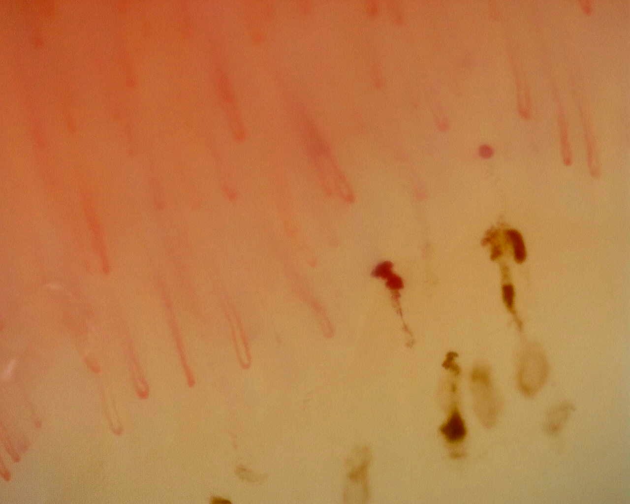
Initial examination
Early scleroderma pattern in a case with Raynaud's phenomenon, swollen hands, and anti-RNP antibodies.
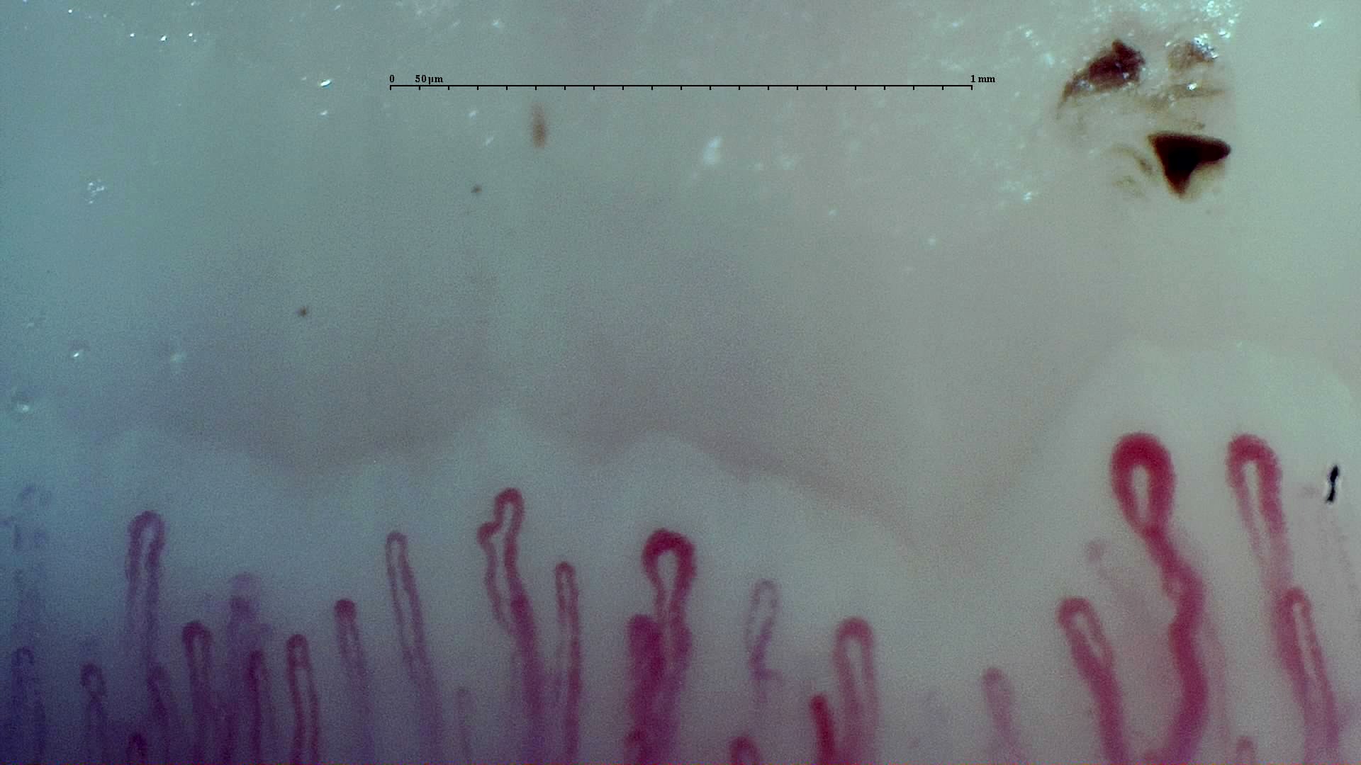
Follow-up
A less pronounced nonspecific pattern on subsequent assessment of the same case.
Pediatric lupus and accumulated damage
Childhood-onset SLE is one of the settings in which capillaroscopy research has added most in recent years [5, 6, 7].
A 2020 systematic review concluded that evidence remained scarce and heterogeneous, although it suggested hemorrhages and abnormal morphology in some patients, with a scleroderma pattern reported in only a minority of series [5]. Shortly afterward, the case-control cohort by Schonenberg-Meinema et al showed more giant capillaries, abnormal shapes, pathological hemorrhages, and pericapillary extravasations than in healthy controls. A scleroderma pattern was present in 17.1% of patients, despite no symptoms of systemic sclerosis [6].
Longitudinal follow-up further refined interpretation. In a subsequent study, the scleroderma pattern was associated with greater accumulated damage, but not progression to systemic sclerosis during follow-up [7]. In practice, markedly abnormal capillaroscopy in pediatric lupus should lead to closer monitoring and may inform prognostic discussions, but should not change the underlying diagnosis while the clinical phenotype remains SLE [5, 6, 7].
| Scenario | What capillaroscopy suggests | What it does not establish alone |
|---|---|---|
| cSLE with abundant hemorrhages | May be associated with greater activity or nephritis [6] | A specific flare; the SLE Disease Activity Index (SLEDAI) and organ findings must be integrated |
| cSLE with a scleroderma pattern | A microvascular phenotype of interest and potentially greater accumulated damage [7] | Automatic progression to systemic sclerosis |
| cSLE without major abnormalities | Does not exclude activity or future involvement [5, 6, 7] | A guaranteed benign course |
cSLE, childhood-onset SLE.
PAH and other clinical questions
PAH is the SLE complication for which capillaroscopy has added the most value to date, although the evidence remains far less developed than in systemic sclerosis [9, 10].
In a retrospective cohort with hemodynamic confirmation, Donnarumma et al reported that a scleroderma pattern was associated with a more than sixfold increase in the relative risk of PAH [9]. A 2023 expert consensus and systematic review on PAH risk factors in SLE included the scleroderma pattern among clinically relevant screening factors, alongside low diffusing capacity for carbon monoxide (DLCO), pericardial effusion, and other markers [10]. The evidence base remains small, however. Its practical value is to raise suspicion, without replacing echocardiography, DLCO, or catheterization when indicated.
Beyond PAH, the literature is less consistent. Some observations suggest links with digital lesions, nephritis, or activity, and there are also disputed data concerning cognitive impairment, Jaccoud's arthropathy, or serum complement C3 and C4 levels [2, 6, 11, 12]. Requests for capillaroscopy in SLE should therefore state the clinical question as explicitly as possible. “Looking for abnormalities” is a weak indication. “SLE with new Raynaud's phenomenon,” “SLE with anti-RNP and swollen hands,” or “SLE with suspected PAH” provide clear reasons for examination.
Insufficient evidence / not found. There is no solid basis for serial capillaroscopy in every patient with SLE as a universal marker of activity or damage.
Reporting without overstating findings
In SLE, reporting style is almost as important as the image. A cautious report describes and contextualizes; a poor report turns weak signals into falsely firm conclusions [1, 2, 12].
- Describe specific domains: hemorrhages, dilations, giant capillaries, density, and architecture.
- State explicitly when the pattern is nonspecific.
- Mention whether the clinical context includes Raynaud's phenomenon, anti-U1-RNP, or suspected PAH.
- Avoid phrases such as “capillaroscopy diagnostic of SLE” or “scleroderma overlap confirmed by the image.”
- If a scleroderma pattern is present, use a conditional conclusion: “findings warranting correlation with clinical features and serology.”
Short cases
Case 1. SLE with Raynaud's phenomenon and anti-U1-RNP
A young woman with SLE, a one-year history of Raynaud's phenomenon, and anti-U1-RNP has dilations, hemorrhages, and some giant capillaries without clear capillary loss. Capillaroscopy strengthens suspicion of a vascular overlap phenotype and supports review of the lungs, hands, and follow-up, but is insufficient to reclassify the disease as systemic sclerosis [3, 8, 10].
Case 2. Stable SLE without vascular symptoms
A patient with low disease activity, no Raynaud's phenomenon, and no digital lesions has mild tortuosity and a punctate traumatic hemorrhage on capillaroscopy. The report should remain conservative: nonspecific findings without evidence of relevant microangiopathy. In this setting, the test adds little and does not justify treatment changes [1, 2, 12].
FAQ
Is there a capillaroscopic pattern specific to SLE?
No validated specific pattern exists. Normal examinations or nonspecific abnormalities are most common [2, 4, 12].
When is capillaroscopy most useful in SLE?
Particularly with Raynaud's phenomenon, anti-U1-RNP, digital lesions, suspected PAH, or uncertainty about overlap [1, 3, 8, 9, 10].
Does a scleroderma pattern in SLE mean overlap with systemic sclerosis?
Not necessarily. It requires clinical and serological correlation, but does not equal confirmed overlap [3, 7].
Can capillaroscopy monitor SLE activity?
It may provide signals of vascular activity in some cohorts, but is not validated as an isolated monitoring biomarker [2, 4, 6, 11, 12].
Glossary
- Nonspecific pattern
- A combination of abnormal findings that does not fit a well-defined pattern and cannot, on its own, identify a specific connective tissue disease.
- Anti-U1-RNP
- An autoantibody associated with MCTD and some SLE phenotypes with Raynaud's phenomenon, PAH, or overlap features.
- Scleroderma pattern
- A combination of giant capillaries, hemorrhages, capillary loss, and neoangiogenesis resembling the systemic sclerosis spectrum, although it may occur in other diseases.
- Vascular activity
- Clinical and morphological expression of microvascular damage or inflammation; no single validated capillaroscopic threshold defines it in SLE.
References
- Smith V, Ickinger C, Hysa E, Snow M, Frech T, Sulli A, et al. Nailfold capillaroscopy. Best Pract Res Clin Rheumatol. 2023;37(1):101849. doi: 10.1016/j.berh.2023.101849. PMID: 37419757.
- Cutolo M, Melsens K, Wijnant S, Ingegnoli F, Thevissen K, De Keyser F, et al. Nailfold capillaroscopy in systemic lupus erythematosus: A systematic review and critical appraisal. Autoimmun Rev. 2018;17(4):344-352. doi: 10.1016/j.autrev.2017.11.025. PMID: 29427827.
- Lambova SN. Scleroderma-like capillaroscopic pattern in SLE is not a sign of overlap syndrome in both adults and children. Lupus Sci Med. 2022;9(1):e000749. DOI: 10.1136/lupus-2022-000749. PMID: 35851262.
- Zhao T, Lin FA, Chen HP. Pattern of nailfold capillaroscopy in patients with systemic lupus erythematosus. Arch Rheumatol. 2020;35(4):568-574. DOI: 10.46497/ArchRheumatol.2020.7763. PMID: 33758813.
- Schonenberg-Meinema D, Melsens K, Nassar-Sheikh Rashid A, Cutolo M, Kuijpers TW, van den Berg JM, et al. Capillaroscopy in childhood-onset systemic lupus erythematosus: a first systematic review. Clin Exp Rheumatol. 2020;38(2):350-354. DOI: 10.55563/clinexprheumatol/azl2bj. PMID: 31858964.
- Schonenberg-Meinema D, Bergkamp SC, Nassar-Sheikh Rashid A, van der Aa LB, de Bree GJ, Ten Cate R, et al. Nailfold capillary abnormalities in childhood-onset systemic lupus erythematosus: a cross-sectional study compared with healthy controls. Lupus. 2021;30(5):818-827. doi: 10.1177/0961203321998750. PMID: 33657918.
- Schonenberg-Meinema D, Bergkamp SC, Nassar-Sheikh Rashid A, Gruppen MP, Middelkamp-Hup MA, Armbrust W, et al. Nailfold capillary scleroderma pattern may be associated with disease damage in childhood-onset systemic lupus erythematosus: important lessons from longitudinal follow-up. Lupus Sci Med. 2022;9(1):e000572. doi: 10.1136/lupus-2021-000572. PMID: 35140136.
- Chebbi PP, Goel R, Ramya J, Gowri M, Herrick A, Danda D. Nailfold capillaroscopy changes associated with anti-RNP antibodies in systemic lupus erythematosus. Rheumatol Int. 2022;42(8):1355-1361. doi: 10.1007/s00296-021-04894-4. PMID: 34076720.
- Donnarumma JFS, Ferreira EVM, Ota-Arakaki J, Kayser C. Nailfold capillaroscopy as a risk factor for pulmonary arterial hypertension in systemic lupus erythematosus patients. Adv Rheumatol. 2019;59(1):1. DOI: 10.1186/s42358-018-0045-5. PMID: 30657102.
- Atsumi T, Bae SC, Gu H, Huang WN, Li M, Nikpour M, et al. Risk Factors for Pulmonary Arterial Hypertension in Patients With Systemic Lupus Erythematosus: A Systematic Review and Expert Consensus. ACR Open Rheumatol. 2023;5(12):663-676. doi: 10.1002/acr2.11611. PMID: 37794618.
- Makarem YS, Selim ZI, Ismail S, Imam Mekkawy A, Galal H, El Nouby FH. Nailfold capillaroscopy changes in systemic lupus erythematosus patients: Correlation with disease activity and anti-uridin1-ribonucleoprotein antibodies. Reumatol Clin (Engl Ed). 2025;21(3):501840. doi: 10.1016/j.reumae.2025.501840. PMID: 40316396.
- Ingegnoli F, Ughi N, Riccieri V, Stefanantoni K, Cracowski JL, Gutierrez M, et al. Distribution of nailfold videocapillaroscopy parameters in systemic lupus erythematosus and their association with disease activity: an international blinded case-control analysis on behalf of the EULAR study group on microcirculation in rheumatic diseases. RMD Open. 2025;11(3):e005772. DOI: 10.1136/rmdopen-2025-005772. PMID: 40921627.