Among inflammatory myopathies, capillaroscopy is most useful in dermatomyositis and, to a lesser extent, antisynthetase syndrome and overlap syndromes. In these phenotypes, it may show a scleroderma-like pattern, correlate with activity, and change with treatment. In immune-mediated necrotizing myopathy (IMNM) and inclusion body myositis (IBM), however, evidence on capillaroscopic findings is scarce or virtually absent [1, 2, 3, 4, 5, 6, 7, 8, 9, 10, 11, 12].
- In current practice, simply distinguishing “polymyositis” from “dermatomyositis” is insufficient; capillaroscopic interpretation depends on the phenotype, serological profile, and systemic context [1, 2, 3, 4].
- Dermatomyositis has the highest frequency of abnormal findings: dilations, giant capillaries, hemorrhages, capillary loss, and scleroderma-like patterns are clearly more common than in other subgroups [5, 6, 7, 8, 9].
- In the systematic review by Flatley et al, increased vessel diameter was present in 64.5%, reduced vascularity in 57.5%, and a scleroderma-spectrum pattern in 45.1% of patients with adult-onset dermatomyositis [7].
- Capillaroscopy in dermatomyositis is more dynamic than previously thought: several cohorts show improvements in hemorrhages, severity scores, and density after immunosuppression [5, 8, 11].
- Antisynthetase syndrome may show abnormalities, but these are usually less pronounced than in dermatomyositis and are not necessarily associated with Raynaud's phenomenon or interstitial lung disease (ILD) [9, 12].
- In IMNM and IBM, the diagnostic value of capillaroscopy is limited; striking findings should prompt reassessment for overlap or misclassification [1, 2, 3, 4, 5].
Learning path
If you suspect dermatomyositis or an inflammatory myopathy with a microvascular component, this unit explains how to interpret a scleroderma-like pattern outside systemic sclerosis. Then review autoantibodies, anti-MDA5, and prognostic value to refine interpretation by serological subgroup [1, 2, 3, 4, 5, 6, 7, 8, 9, 10, 11, 12].
Where capillaroscopy fits in myopathies
Capillaroscopy is not part of the myositis classification criteria, but it adds value in phenotype characterization and differential diagnosis when the clinical picture is not yet well established [1, 2, 3, 4].
This matters because inflammatory myopathies are a heterogeneous group. The 2017 ACR/EULAR classification (American College of Rheumatology / European Alliance of Associations for Rheumatology) remains a useful foundation, but does not fully capture the serological and phenotypic diversity encountered today [1, 2, 3]. In this context, capillaroscopy is most useful as a marker of associated vasculopathy or microangiopathy, rather than as a standalone test that classifies the disease [1, 2, 4].
The structured review by the European Alliance of Associations for Rheumatology (EULAR) microcirculation group summarizes the current position: available data are more consistent in dermatomyositis and less robust in other subtypes. Terminology should also align with that used in systemic sclerosis to avoid arbitrary descriptions [5]. Readers should use recognizable language rather than inventing myositis-specific categories.
| Clinical question | What capillaroscopy can add | Main limitation |
|---|---|---|
| Does the phenotype more closely resemble dermatomyositis? | More dilation, giant capillaries, hemorrhages, and scleroderma-like patterns [4, 5, 6, 7, 8] | Does not replace skin, muscle, or autoantibody assessment, or biopsy |
| Is there microvascular activity that can be followed? | Hemorrhages, density, and quantitative scores may change with treatment [5, 8, 11] | Universally accepted thresholds are lacking |
| Is this antisynthetase syndrome or overlap? | A nonspecific or less pronounced scleroderma-like pattern may be present [9] | Considerable heterogeneity across cohorts |
| Is it useful in IMNM or IBM? | Very limited or virtually absent evidence [1, 4, 5] | Risk of overinterpretation |
Dermatomyositis: the phenotype in which capillaroscopy adds the most
Dermatomyositis is the inflammatory myopathy in which capillaroscopy is most useful in practice. It is where the test shows the most quantitative and qualitative abnormalities and where the literature is most consistent [4, 5, 6, 7, 8].
In the systematic review by Flatley et al, which included 346 adults with dermatomyositis, capillaroscopy found increased vessel diameter in 64.5%, reduced vascularity in 57.5%, and a scleroderma-spectrum pattern in 45.1% [7]. These figures confirm what earlier cohorts had suggested: microvascular abnormalities are common in dermatomyositis and usually manifest as dilation, capillary loss, hemorrhages, and disorganization [4, 5, 6, 7, 8].
The Japanese cohort by Kubo et al reinforced this observation by comparing myopathy phenotypes. Capillaroscopic abnormalities occurred in 55.7% overall, but were much more frequent in dermatomyositis than in polymyositis: 65.4% versus 27.8%. The frequency reached 87.5% in anti-MDA5-positive and 88.9% in anti-TIF1gamma-positive patients [6]. The multicenter series by Torres-Ruiz et al added an important qualification: a truly normal pattern was uncommon, at around 9% of cases, and the scleroderma pattern was mainly seen in dermatomyositis and overlap syndromes [8].
Xu's review of 76 patients with dermatomyositis summarizes the typical profile: 73.7% had abnormal capillaroscopy, with greater severity than in antisynthetase syndrome but less than in systemic sclerosis [9]. Overall, when an inflammatory myopathy is suspected and giant capillaries, hemorrhages, and rarefaction are present, the findings favor dermatomyositis or an overlap syndrome with a strong microvascular component [4, 5, 6, 7, 8].
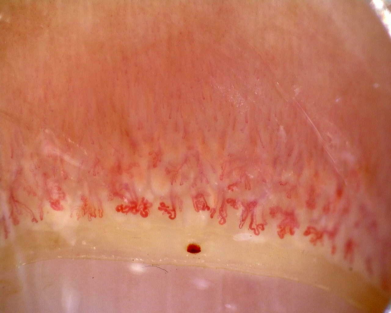
| Capillary domain | Dermatomyositis | Practical comment |
|---|---|---|
| Dilation / giant capillaries | Frequent [5, 6, 7, 8] | Carry more weight when accompanied by hemorrhages and capillary loss |
| Capillary loss / avascular areas | Common and associated with greater vascular burden [5, 6, 7, 8] | An isolated image with few capillaries is not always interpretable |
| Scleroderma-like pattern | Much more frequent than in other subtypes [4, 5, 6, 7, 8] | Does not automatically imply systemic sclerosis |
| Disorganization / neoangiogenesis | May occur in active or complex cases [4, 7, 8] | Evidence on associated cancer remains limited |
Antisynthetase syndrome and overlap syndromes
Antisynthetase syndrome may include Raynaud's phenomenon, mechanic's hands, telangiectasias, or overlap features, but its capillaroscopic findings are usually less pronounced than those of dermatomyositis [1, 4, 6, 9, 12].
Sebastiani's international study analyzed 190 patients with antisynthetase syndrome and detected abnormalities in 62.1%: giant capillaries in 31.6%, avascular areas in 20%, and a scleroderma-like pattern in 35.3% [10]. This is a meaningful frequency, but remains below that observed in many dermatomyositis cohorts; the pattern was also not significantly associated with Raynaud's phenomenon [10].
This is clinically relevant. In systemic sclerosis, the scleroderma pattern tends to be interpreted as a consequence of structural microangiopathy in a setting dominated by vasculopathy. In antisynthetase syndrome and other inflammatory myopathies, the pathophysiology appears more inflammatory and less closely tied to a pure ischemia–reperfusion model [4, 6, 9]. This difference explains why parallels with systemic sclerosis require caution.
The more recent study by Mugii et al explored this heterogeneity by comparing six anti-ARS subtypes in 66 patients. Overall, dilations, fewer visible capillaries, and hemorrhages were the most frequent findings. Complex abnormal shapes, disorganization, and capillary loss were rare, and the anti-KS subtype had a clearly milder microvascular profile than the others [12]. The article's abstract still uses the classical term capillary ramifications [12], which these units include within the more current category of abnormal shapes.
In overlap syndromes, particularly scleromyositis, capillaroscopy may be more informative because microvascular damage and scleroderma features coexist. However, many cohorts combine phenotypes and treatments, so specific percentages should be regarded as indicative rather than definitive [1, 2, 3, 4, 8, 9].
| Phenotype | Expected utility* | Practical interpretation |
|---|---|---|
| Dermatomyositis | High [5, 6, 7, 8] | Capillaroscopy adds diagnostic and follow-up information |
| Antisynthetase syndrome | Intermediate [6, 10, 12] | May be useful, but the pattern is usually less pronounced and less specific |
| Scleromyositis / overlap | Intermediate to high [1, 4, 8, 9] | Interpret in the context of scleroderma features and autoantibodies |
*Refers to the information provided by capillaroscopy.
Phenotypes in which capillaroscopy is less useful
Capillaroscopic findings should not carry the same weight in every inflammatory myopathy. The most frequent error is extrapolating what is known about dermatomyositis to IMNM or IBM [1, 2, 3, 4, 5].
In IMNM, the literature is very limited and reported findings are scattered, often based on small numbers or overlap cases [1, 4, 5]. In IBM, evidence on capillaroscopic utility is virtually absent [1, 2, 3, 4]. This does not mean that abnormalities can never occur, but there is no solid basis for making capillaroscopy a central tool in these phenotypes.
Insufficient evidence / not found. There is no robust support for using capillaroscopy as a routine biomarker in IMNM or sporadic IBM.
Practical reporting
A useful report in inflammatory myopathies should go beyond the label “dermatomyositis pattern.” Describe individual domains first, then explain whether the overall findings resemble a scleroderma-like pattern in the context of an inflammatory myopathy [4, 8].
- State whether the images are evaluable and how many fingers could be assessed.
- Describe density, giant capillaries, hemorrhages, avascular areas, and neoangiogenesis.
- If you report a scleroderma-like pattern, explain its basis and how it differs from a typical systemic sclerosis phase.
- Relate the findings to the suspected phenotype, such as dermatomyositis, antisynthetase syndrome, or overlap.
- State the limitations: a nonspecific pattern neither confirms nor excludes myositis.
Short cases
Case 1. Proximal weakness and a heliotrope rash
A patient with proximal weakness, Gottron's papules, and elevated creatine kinase (CK) has giant capillaries, hemorrhages, and mild capillary loss. Capillaroscopy does not replace myositis classification, but supports a dermatomyositis phenotype and provides a baseline for follow-up [1, 4, 5, 6, 7, 8].
Case 2. Antisynthetase syndrome with mechanic's hands
In an anti-Jo1-positive woman with ILD and mechanic's hands, capillaroscopy shows dilations and some abnormal shapes without marked rarefaction. This fits the expected findings in antisynthetase syndrome: abnormalities may occur, but are less pronounced and less defining than in dermatomyositis [6, 10].
FAQ
Which inflammatory myopathy shows the most capillaroscopic abnormalities?
Dermatomyositis, followed by antisynthetase syndrome and other phenotypes [4, 5, 6, 7, 8, 9].
Can capillaroscopy diagnose myositis?
Not on its own. It helps characterize the phenotype and supports clinical suspicion, but does not replace muscle, skin, serological, or histological assessment [1, 2, 3, 4].
Can capillaroscopy normalize with treatment?
Yes, particularly in dermatomyositis. Hemorrhages may improve or disappear, and some scores may improve after immunosuppression [5, 8, 11].
Does a scleroderma-like pattern in dermatomyositis mean systemic sclerosis?
No. It indicates similar microangiopathy, but its clinical meaning depends on the overall myositis phenotype and serology [4, 5, 6, 7, 8].
Glossary
- Idiopathic inflammatory myopathies
- A heterogeneous group of systemic diseases with predominantly muscular involvement and diverse clinical, serological, and pathological phenotypes.
- Dermatomyositis
- A myositis phenotype with characteristic skin involvement and the condition in which capillaroscopy adds the most information.
- Antisynthetase syndrome
- A phenotype associated with anti-aminoacyl-tRNA synthetase autoantibodies, ILD, arthritis, mechanic's hands, and variable Raynaud's phenomenon.
- IMNM
- Immune-mediated necrotizing myopathy, a subtype in which capillaroscopy currently has limited utility.
- Scleroderma-like pattern
- A set of capillaroscopic findings resembling the systemic sclerosis pattern, without necessarily implying that the disease is present.
References
- Selva-O'Callaghan A, Pinal-Fernández I, Trallero-Araguás E, Milisenda JC, Grau-Junyent JM, Mammen AL. Classification and management of adult inflammatory myopathies. Lancet Neurol. 2018;17(9):816-828. doi: 10.1016/S1474-4422(18)30254-0. PMID: 30129477.
- Lundberg IE, Tjärnlund A, Bottai M, Werth VP, Pilkington C, de Visser M, et al. 2017 European League Against Rheumatism/American College of Rheumatology Classification Criteria for Adult and Juvenile Idiopathic Inflammatory Myopathies and Their Major Subgroups. Arthritis Rheumatol. 2017;69(12):2271-2282. doi: 10.1002/art.40320. PMID: 29106061.
- Zanframundo G, Selva-O'Callaghan A, González-Gay MÁ, Montecucco C, Cavagna L. Issues in the classification of myositis patients: an ongoing process. Clin Exp Rheumatol. 2024 Feb;42(2):225-228. doi: 10.55563/clinexprheumatol/8u8p8x. Epub 2024 Feb 13. PMID: 38372711.
- Pauling JD, Christopher-Stine L. The aetiopathogenic significance, clinical relevance and therapeutic implications of vasculopathy in idiopathic inflammatory myopathy. Rheumatology (Oxford). 2021 Apr 6;60(4):1593-1607. doi: 10.1093/rheumatology/keaa816. Erratum in: Rheumatology (Oxford). 2021 Aug 2;60(8):3960. doi: 10.1093/rheumatology/keab431. PMID: 33458769.
- Piette Y, Reynaert V, Vanhaecke A, Bonroy C, Gutermuth J, Sulli A, et al. Standardised interpretation of capillaroscopy in autoimmune idiopathic inflammatory myopathies: A structured review on behalf of the EULAR study group on microcirculation in Rheumatic Diseases. Autoimmun Rev. 2022 Jun;21(6):103087. doi: 10.1016/j.autrev.2022.103087. Epub 2022 Apr 12. PMID: 35421608.
- Kubo S, Todoroki Y, Nakayamada S, Nakano K, Satoh M, Nawata A, et al. Significance of nailfold videocapillaroscopy in patients with idiopathic inflammatory myopathies. Rheumatology (Oxford). 2019 Jan 1;58(1):120-130. doi: 10.1093/rheumatology/key257. PMID: 30169697.
- Flatley EM, Collins D, Lukowiak TM, Miller JH. Nailfold microscopy in adult-onset dermatomyositis in association with myositis antibodies. Arch Dermatol Res. 2024 Nov 20;317(1):34. doi: 10.1007/s00403-024-03521-z. PMID: 39565371.
- Torres-Ruiz J, Pinal-Fernandez I, Selva-O'Callaghan A, Campbell B, Muñoz-Braceras S, Mejía-Domínguez NR, et al. Nailfold capillaroscopy findings of a multicentric multi-ethnic cohort of patients with idiopathic inflammatory myopathies. Clin Exp Rheumatol. 2024 Feb;42(2):367-376. doi: 10.55563/clinexprheumatol/l9gudh. Epub 2024 Mar 14. PMID: 38488092.
- Xu H, Qian J. The role of nailfold video-capillaroscopy in the assessment of dermatomyositis. Rheumatology (Oxford). 2025 May 1;64(5):2987-2994. doi: 10.1093/rheumatology/keae677. PMID: 39689020.
- Sebastiani M, Triantafyllias K, Manfredi A, González-Gay MA, Palmou-Fontana N, Cassone G, et al. Nailfold Capillaroscopy Characteristics of Antisynthetase Syndrome and Possible Clinical Associations: Results of a Multicenter International Study. J Rheumatol. 2019 Mar;46(3):279-284. doi: 10.3899/jrheum.180355. Epub 2018 Nov 15. PMID: 30442823.
- Mugii N, Hamaguchi Y, Horii M, Fushida N, Ikeda T, Oishi K, et al. Longitudinal changes in nailfold videocapillaroscopy findings differ by myositis-specific autoantibody in idiopathic inflammatory myopathy. Rheumatology (Oxford). 2023 Mar 1;62(3):1326-1334. doi: 10.1093/rheumatology/keac401. PMID: 35866689.
- Mugii N, Hamaguchi Y, Fushida N, Fujii K, Nishio J, Kudo K, et al. Distinct Nailfold Videocapillaroscopy Findings in Patients With Anti-Aminoacyl-tRNA Synthetase Antibodies. J Dermatol. 2026;53(2):318-322. doi: 10.1111/1346-8138.70081. PMID: 41312673.
- McClellan N, Chen L, Matossian S, et al. Nailfold capillary phenotypes distinguish juvenile myositis subtypes and associate with disease activity. Clin Transl Med. 2026;16(9):e70789. doi: 10.1002/ctm2.70789. PMID: 42681940.