Equipment and devices for capillaroscopy
Choosing the right equipment shapes the quality of the entire process: which structures you will be able to see, which measurements will be comparable between visits, and how much confidence your report will carry. Videocapillaroscopy remains the reference standard for documentation and follow-up, but not every clinical setting needs the same hardware level or the same budget [1, 2, 3].
- Videocapillaroscopy remains the reference standard when patients need to be measured, archived, and followed longitudinally [1, 2, 6].
- The commercial magnification number is not enough to choose a device; real field of view, sharpness, and calibration matter more [1, 2, 5].
- Universal Serial Bus (USB) microscopes and other low-cost options may be useful for screening or gradual implementation, but they should not be assumed equivalent for all parameters [3, 5].
- Nailfold dermoscopy may have value as an initial filter in Raynaud's phenomenon, not as a universal substitute for a structured capillaroscopic study [2, 4].
- Software, image archiving, and traceability matter as much as optics if the aim is to compare studies over time [2, 6, 7, 8, 9].
Learning path
If you are deciding which equipment to start with, this unit explains what each option adds and in which context it makes sense to use it. Afterward, it will be easier to review acquisition and quality to decide how to capture useful images with the hardware available to you.
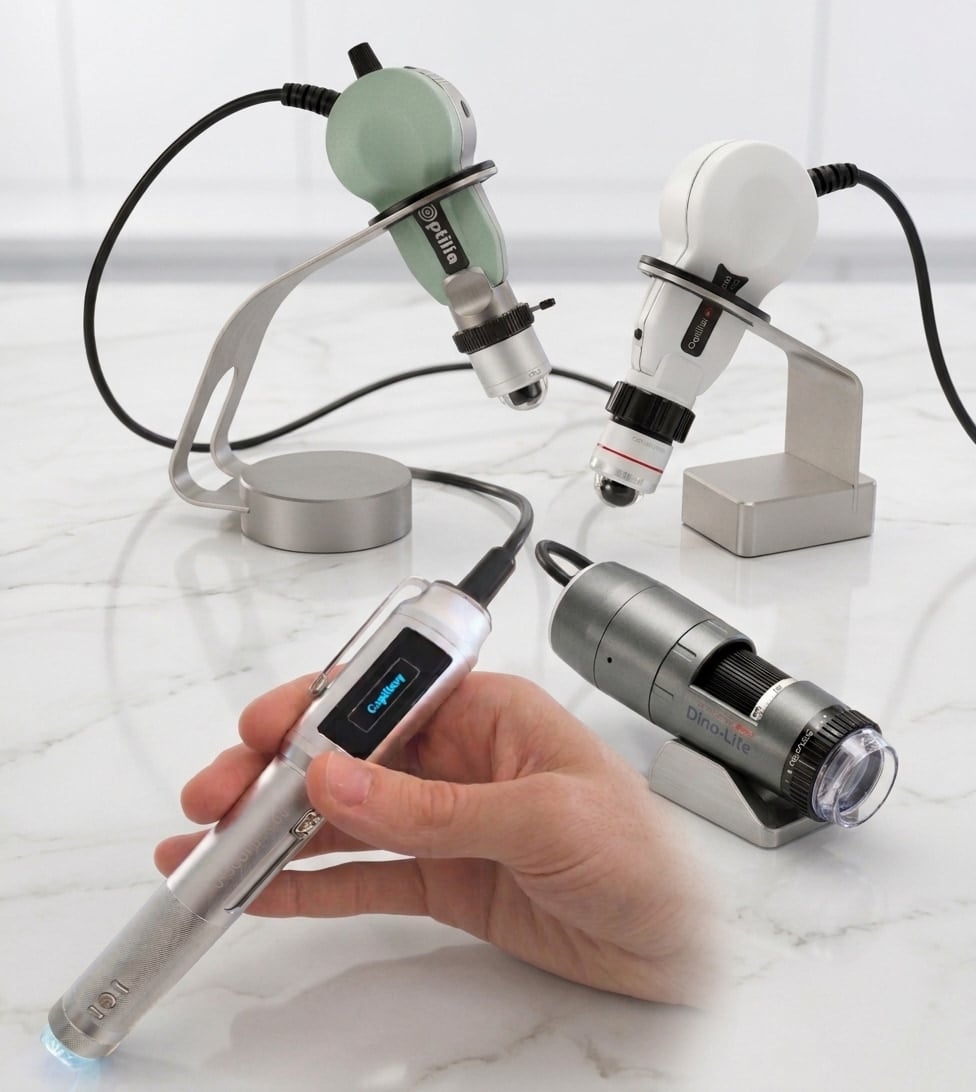
Device overview
The right question is not which device is the "most advanced", but which clinical decision you need to support and what level of traceability is required.
| Device type | Reasonable use | Main strength | Main limitation |
|---|---|---|---|
| Videocapillaroscope | Structured diagnosis, follow-up, and advanced teaching | Best integration of optics, calibration, archiving, and measurement [1, 2] | Higher upfront cost and more demanding implementation |
| Stereomicroscope | Teaching, laboratories, or settings with previous optical equipment | Good availability in some centers | Less clinical ergonomics and weaker digital integration [2, 5] |
| Low-cost USB microscope | Low-budget clinic, pilot projects, or initial learning | Accessibility and portability | Uneven performance depending on the analyzed parameter [3, 5] |
| Digital dermoscope | Rapid screening in Raynaud's phenomenon or dermatology clinics | Widely available and easy to use [2, 4] | Not the best option for fine quantification or detailed follow-up [2, 4] |
| Smartphone with optical adapter | Teleorientation, basic training, or very preliminary documentation | Very low cost and maximum portability | High heterogeneity between configurations and limited standardization [2] |
Recent methodological reviews continue to place videocapillaroscopy as the reference technique because it does not simply "see better": it enables comparable measurements, longitudinal follow-up, and the use of quantitative or semi-automated tools [1, 2, 6]. This is especially relevant when the aim is to differentiate primary Raynaud's phenomenon from scleroderma-spectrum microangiopathy, document progression, or support a clinical decision.
This does not mean that every simpler system lacks value. It means that simpler systems should be used with more modest and explicit objectives. In the pilot study comparing videocapillaroscopy with low-cost USB microscopy, some simple features, such as capillary width or identification of well-defined capillaries, translated reasonably well; by contrast, density and part of the morphological analysis performed worse with the cheaper system [3].
Nailfold dermoscopy occupies an intermediate space. The prospective multicenter VASCUL-R trial showed that a normal dermoscopy, defined by the absence of four simple findings, predicted a normal videocapillaroscopy with a positive predictive value of 100%; however, its sensitivity was not sufficient to make it a substitute for the full study [4]. Translated into clinic: it works well as a rapid filter, not as a single rule to close the case when clinical suspicion is high.
| Feature | Smart G-Scope | Dino-Lite Capillaryscope 200 Pro | Optilia Digital Capillaroscope | Inspectis Digital Capillaroscope |
|---|---|---|---|---|
| Light | White LED with on/off control | White LED | Integrated RingLight with 12 ultrabright white LEDs and intensity control | Integrated pure white LED light |
| Magnification | 250x for nailfold capillaroscopy | ~50x and ~200x | 100x, 200x, and 300x | 100x, 200x, and 300x |
| Field of view at 200x | ~2.35 x 1.32 mm | ~1.85 x 1.45 mm | ~1.7 x 1.3 mm | ~1.8 x 1.35 mm |
| Resolution | 3.5 megapixels | 1.3 megapixels | 5 megapixels | 5 megapixels |
| Cost | EUR 750-800 | EUR 750-800 | >EUR 6,000 | >EUR 6,000 |
Key technical criteria
Many purchasing errors come from focusing on a single attribute. Real practice requires assessing the combination of optics, ergonomics, and software.
Field of view before nominal magnification
Clinical capillaroscopy usually works around 200x magnification because that range provides sufficient detail without completely sacrificing the analyzed surface [1, 2, 5]. The problem is that two manufacturers can advertise "similar" magnification and provide very different fields of view. To decide, it is more useful to ask how many millimeters of nailfold bed fit into a truly usable image and whether that image includes a reliable scale [2, 5].
| Criterion | What to require | Why it matters |
|---|---|---|
| Useful field of view | Enough to count capillaries per mm without losing sharpness [1, 2] | Avoids insufficient sampling and excessive captures |
| Real optical quality | Sharp capillaries, low noise, and reasonable reflection control [2, 5] | Electronic resolution does not correct poor optics |
| Calibration | Stable or verifiable scale for measurements [2, 6] | Without calibration, density and diameters lose comparability |
| Ergonomics | Stable head, comfortable grip, and support that limits compression | Reduces pressure artifacts and operator fatigue |
| Software and archiving | Finger labeling, retrieval by date, and simple export [2, 7, 8, 9] | Without traceability, serious follow-up is impossible |
Light, polarization, and contact
Illumination and reflection control influence image readability as much as the camera. Polarization can help, but it should not be purchased as if it automatically guaranteed quality. Depending on optics, phototype, and the amount of oil or gel, it can also modify the background color or reduce the useful contrast of fine capillaries [2, 5].
Something similar happens with contact between the head and the skin. A very bulky or poorly balanced device forces more pressure and increases the probability of capillary collapse, especially in cold hands or small nailfold beds. This limitation rarely appears in the commercial specification sheet, but it appears in daily practice.
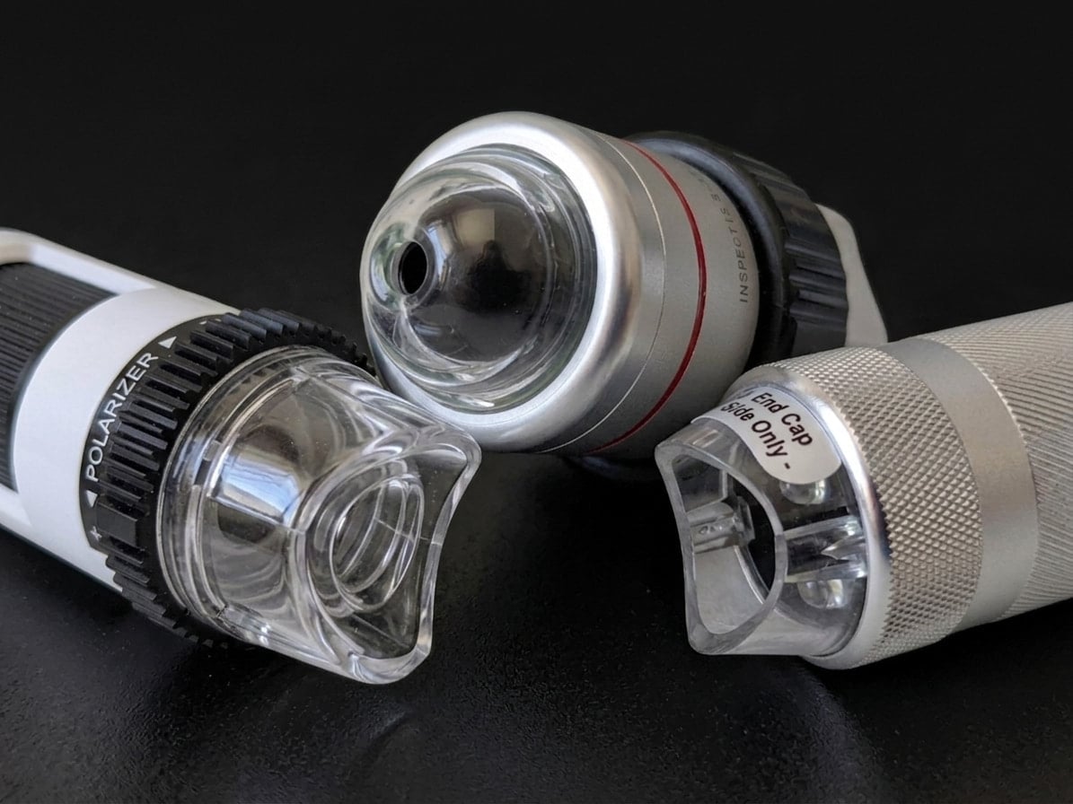
The main models have two types of heads: concave heads, which allow greater support but may be more difficult in certain younger or thinner patients depending on finger size, and convex heads, which require somewhat more learning but adapt better to different populations, including pediatric patients, because they need less support surface.
Software is not an accessory
Software determines whether an image can become clinical data. It should allow fingers to be named, date and laterality to be saved, previous studies to be retrieved, calibration to be checked, and files to be exported without losing context [2, 6, 7, 8, 9]. The minimum core domains proposed for capillaroscopy reports in 2024 point precisely in that direction: capturing the image is not enough; the process and result must be recorded so that another person can understand and compare them [7].
This has a very clear practical consequence. If two devices generate acceptable images but one does not allow those images to be identified, ordered by finger, or retrieved quickly, that device will perform worse in longitudinal practice even if the optics are similar. In capillaroscopy, a chaotic archive ultimately degrades clinical quality as much as a poor lens.
Implementation by clinical context
The most prudent decision is not always to buy the most advanced system. Often it is to implement a system that matches patient volume, team training, and the clinical questions that will actually be answered.
Scenario 1: dedicated Raynaud's or systemic sclerosis clinic
In this setting, dedicated videocapillaroscopy is usually the most reasonable option. Longitudinal follow-up, assessment of progression, and the need to document density, dilations, hemorrhages, or global patterns with homogeneous quality are expected [1, 2, 6]. In this scenario, saving money on software or calibration often becomes costly later because it prevents comparison of studies or teaching complex cases to the rest of the team.
Scenario 2: service with limited budget and low patient volume
If the priority is to discriminate which patients with Raynaud's need referral or a complete study, it may make sense to start with a locally validated USB system or digital dermoscopy, provided the center accepts its limits from the outset [3, 4, 5]. In this situation, the most prudent strategy is to use the simpler device as the first step and maintain a clear referral pathway for doubtful cases or abnormal findings.
Scenario 3: teaching or gradual implementation
To learn how to locate the distal row, recognize artifacts, and become familiar with capillary anatomy, a simple system may be sufficient at the start. The important point is not to confuse that learning phase with full diagnostic equivalence. A basic device can teach spatial orientation, but it is not necessarily enough to build a complex follow-up clinic.
| Context | Reasonable solution | What to monitor |
|---|---|---|
| Specialized clinic with follow-up | Dedicated videocapillaroscope | This is where calibration, software, and archiving pay off most [1, 2, 6, 7] |
| General clinic with few cases | Locally validated USB system or referral to an expert center | The key is to recognize the limit and avoid overinterpretation [3, 5] |
| Initial Raynaud's screening | Digital dermoscopy | Useful for prioritizing who needs a complete study [4] |
| Teaching and initial training | Simple system with good educational support | Useful for acquiring visual language, not for assuming full technical equivalence |
Purchase pitfalls and local validation
Implementation fails less often because of one isolated optical choice than because of poor definition of the complete clinical workflow.
Common purchasing errors
- Buying based on magnification number without reviewing the real field of view.
- Ignoring whether verifiable calibration exists for linear measurements [2, 6].
- Not checking how studies are named and retrieved by finger and date [2, 7].
- Choosing a device that requires excessive head pressure or produces strong specular glare.
- Assuming that every digital device is automatically valid for follow-up.
What to validate before using it in real clinical practice
Local validation does not need to be a formal trial, but it should answer several simple questions. Can you obtain sharp images in hands with different phototypes? Can the operator move along the distal row without compressing it? Is the scale stable? Can the images be retrieved months later? Do two observers understand which finger and which sector they are viewing? If any answer is "no", the problem is not minor: it directly affects the clinical utility of the study.
In centers starting from scratch, a sensible strategy is to test the equipment in a small series of healthy volunteers and a few known patients, documenting whether density and major findings are consistently legible. This step does not replace published evidence, but it prevents avoidable local implementation errors.
Checklist for buying or implementing equipment
- Define whether the use will be screening, structured diagnosis, or longitudinal follow-up.
- Require demonstration of the real field of view, not only nominal magnification [1, 2].
- Check calibration and image export before completing the purchase [2, 6].
- Ask for examples of complete studies, not only manufacturer-selected images.
- Verify that the software allows finger labeling and visit comparison [2, 7, 8, 9].
- Plan minimum operator training and a referral pathway for uncertain cases.
Short cases
Concrete examples help show why equipment choice modifies clinical performance.
Case 1: Raynaud's in a dedicated clinic
A 29-year-old woman has biphasic Raynaud's phenomenon, positive antinuclear antibodies (ANA), and puffy fingers. If the plan is to compare her across successive visits and document density, hemorrhages, and giant capillaries, a dedicated system with structured archiving is clearly preferable. In this scenario, a simple device may orient the reader, but it falls short for serious follow-up.
Case 2: first screen in a general clinic
A 24-year-old man has mild acrocyanosis, no autoantibodies, and no other systemic signs. Here a simple system or digital dermoscopy may have value as an initial filter. If the image is clearly normal and clinical suspicion is low, immediate escalation to full videocapillaroscopy may not be necessary; if dilations, repeated hemorrhages, or technical doubts appear, referral would be justified [4].
Case 3: implementation error
A service buys a device advertised with high magnification but without finger-based archiving software. Six months later, it has sharp images but cannot ensure which ones correspond to the right fourth finger or compare them reliably with previous studies. The problem is not secondary: that device can produce attractive images and still perform poorly for clinical follow-up.
FAQ
Is the best device always the one with the highest magnification?
No. What matters is the balance between useful detail, field of view, reflection control, and archiving capacity [1, 2, 5].
Can I start with an inexpensive USB microscope?
Yes, in selected contexts. The prudent approach is to use it for limited objectives and locally validate which parameters are consistently legible [3, 5].
Is dermoscopy useful for Raynaud's phenomenon?
It can be a good initial screening tool, but it does not replace standard capillaroscopy when clinical suspicion is high or quantification is needed [4].
Is polarization essential?
No. It can help control reflections, but its usefulness depends on the optical system and the individual patient [2, 5].
What should the software do at minimum?
Save images by finger, preserve temporal traceability, allow export, and facilitate longitudinal comparison [2, 6, 7, 8, 9].
Does it make sense to buy a very complete system if I see few patients?
Only if the center will concentrate cases and follow them in a structured way. Otherwise, it may be more sensible to use a simpler first step and refer complex cases.
Glossary
- Field of view
- Area of nailfold bed visible in a useful image. In clinical practice, it is often more important than the commercial magnification number.
- Calibration
- Known relationship between pixels and real distance, essential for measuring density or diameters in a comparable way.
- Traceability
- Ability to identify, store, and retrieve images by hand, finger, and date for clinical follow-up.
- Screening
- Use of a simple system to decide which patients need a more complete capillaroscopic evaluation.
- Local validation
- Practical confirmation that a device and workflow function in the real setting where they will be used.
References
- Smith V, Ickinger C, Hysa E, Snow M, Frech T, Sulli A, et al. Nailfold capillaroscopy. Best Pract Res Clin Rheumatol. 2023;37(1):101849. doi: 10.1016/j.berh.2023.101849. PMID: 37419757.
- Smith V, Herrick AL, Ingegnoli F, Damjanov N, De Angelis R, Denton CP, et al. Standardisation of nailfold capillaroscopy for the assessment of patients with Raynaud's phenomenon and systemic sclerosis. Autoimmun Rev. 2020;19(3):102458. doi: 10.1016/j.autrev.2020.102458. PMID: 31927087.
- Berks M, Dinsdale G, Marjanovic E, Murray A, Taylor C, Herrick AL. Comparison between low cost USB nailfold capillaroscopy and videocapillaroscopy: a pilot study. Rheumatology (Oxford). 2021;60(8):3862-3867. doi: 10.1093/rheumatology/keaa723. PMID: 33232464.
- Monfort JB, Klejtman T, Lazareth I, Kottler D, Blaise S, Imbert B, et al. Nailfold dermoscopy predicts the absence of a capillaroscopy sclerodermic pattern: The multicentre, prospective VASCUL-R trial. J Eur Acad Dermatol Venereol. 2024;38(10):1982-1987. doi: 10.1111/jdv.19803. PMID: 38251814.
- Karbalaie A, Emrani Z, Fatemi A, Etehadtavakol M, Erlandsson BE. Practical issues in assessing nailfold capillaroscopic images: a summary. Clin Rheumatol. 2019;38(9):2343-2354. doi: 10.1007/s10067-019-04644-9. PMID: 31278512.
- Herrick AL, Berks M, Taylor CJ. Quantitative nailfold capillaroscopy-update and possible next steps. Rheumatology (Oxford). 2021;60(5):2054-2065. doi: 10.1093/rheumatology/keab006. PMID: 33493310.
- El Miedany Y, Ismail S, Wadie M, Müller-Ladner U, Giacomelli R, Liakouli V, et al. Development of a core domain set for nailfold capillaroscopy reporting. Reumatol Clin (Engl Ed). 2024;20(7):345-352. doi: 10.1016/j.reumae.2024.07.003. PMID: 39160005.
- El Miedany Y, Ismail S, Wadie M, Hassan M. Nailfold capillaroscopy: tips and challenges. Clin Rheumatol. 2022;41(12):3629-3640. doi: 10.1007/s10067-022-06354-1. PMID: 36040673.