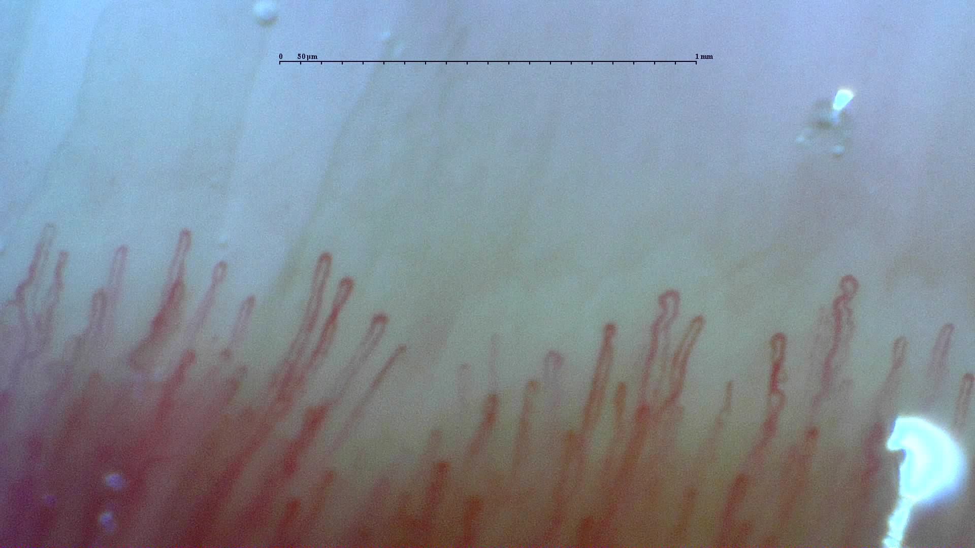
 Normal reference: distal capillary row
A field from the normal reference study, showing the distal row of narrow loops. Use the full study to compare density and shape across fingers.
View the complete studyHow to assess and measure
Describe the morphology in a focused view of the distal row and use a consistent classification if counting altered shapes. Check whether crossing loops are separate vessels and assess apical size independently with calibration. Record density and the surrounding architecture before interpreting shape variation.
Interpretation and related diseases
Some tortuosity can occur as normal variation or in a nonspecific pattern. Isolated winding loops with preserved density and no giants should not be interpreted like a field with capillary loss and complex ramifications. The normal and nonspecific reference studies allow comparison across several fingers.
Read the detailed clinical chapterCommon mistakes
- Calling every crossing or winding loop neoangiogenesis.
- Diagnosing connective tissue disease from tortuosity alone.
- Confusing digital enlargement of the image with true capillary dilation.
Real images and comparisons
Captions distinguish the illustrated finding from the study interpretation. Comparisons include variants, other clinical contexts or artifacts where available.
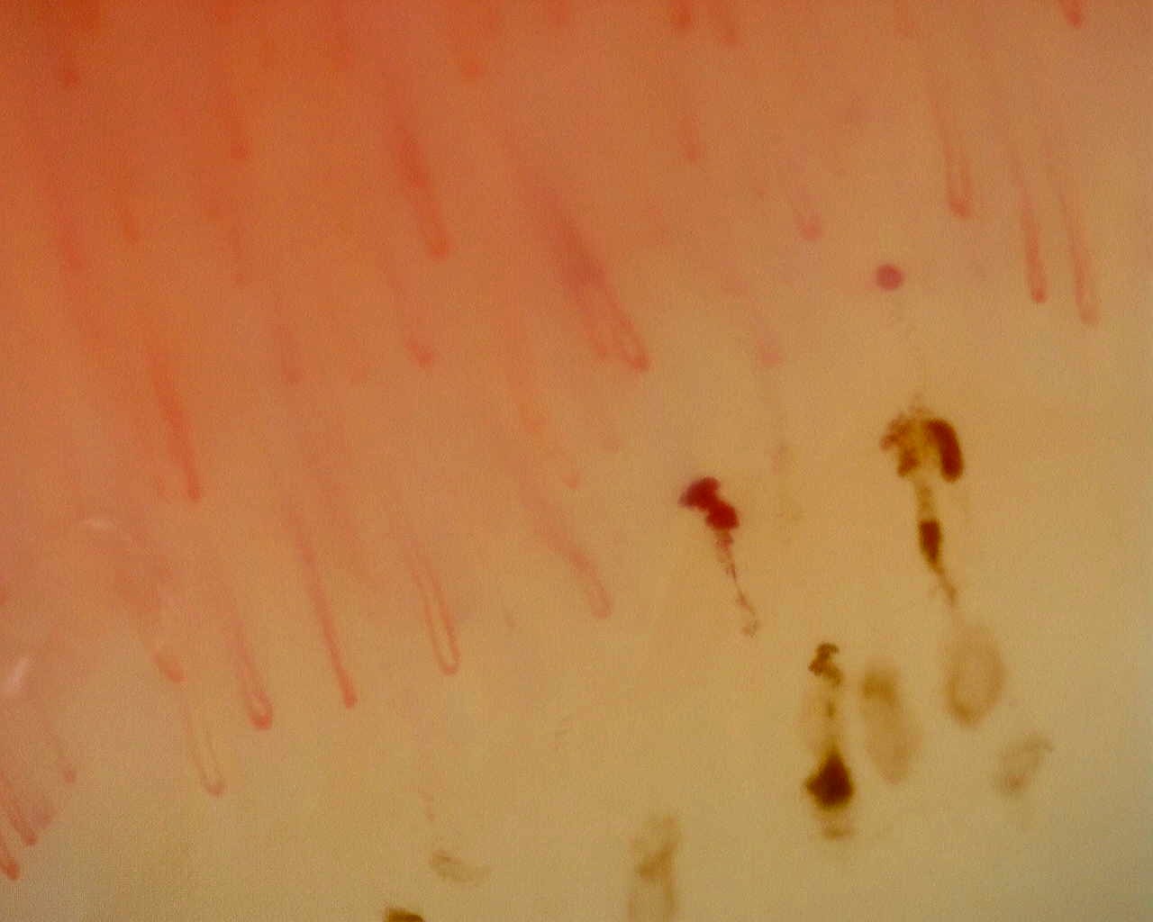 Nonspecific pattern: hemorrhagic field
Dark hemorrhagic deposits alongside capillary loops in the nonspecific reference study. Hemorrhage alone does not define a scleroderma pattern.
View the complete studyComplete cases and studies
Normal Pattern: Representative Case
The complete image set shows a regular distal row, preserved density, absent giant capillaries, and no avascular areas.
Non-Specific Pattern: Representative Case
The study shows preserved density, around 8.8 capillaries/mm, scattered dilated or tortuous loops, and hemorrhages around 0.46/mm, with no giant capillaries and no Cutolo SSc pattern.
References
- Smith V, et al. Standardisation of nailfold capillaroscopy for the assessment of patients with Raynaud’s phenomenon and systemic sclerosis. Autoimmun Rev. 2020;19:102458.
- Cutolo M, et al. Nailfold videocapillaroscopy assessment of microvascular damage in systemic sclerosis. J Rheumatol. 2000;27:155–160.
- Ingegnoli F, et al. Reporting items for capillaroscopy in clinical research on musculoskeletal diseases: a systematic review and international Delphi consensus. Rheumatology. 2021;60:1410–1418.
Scientific advisory board and contributors · Editorial policy