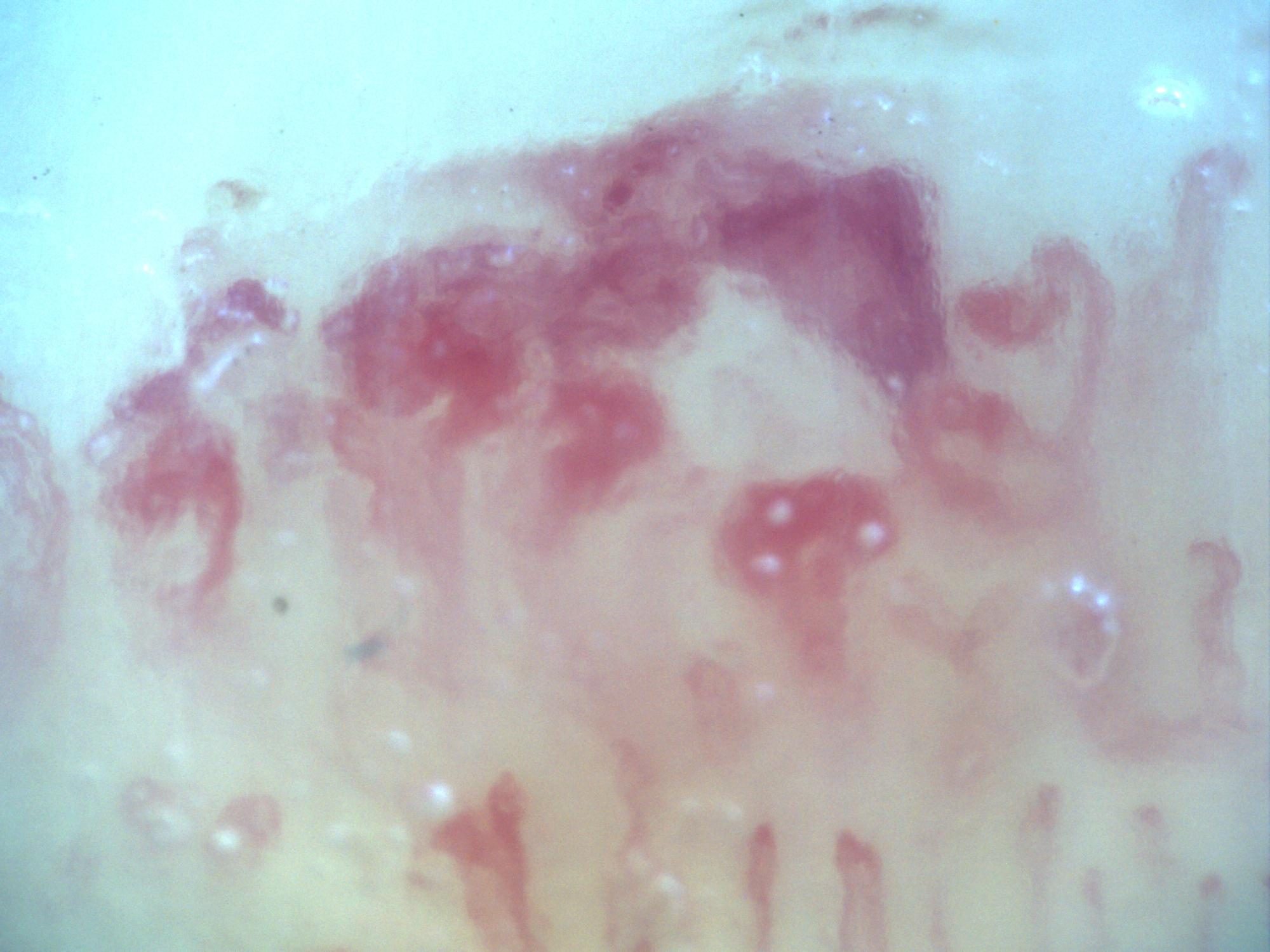
200x capillaroscopy image from systemic sclerosis showing a microvascular hemorrhage with the typical smoke-plume appearance.
How to assess and measure
Document presence, location and distribution across fingers. If quantifying hemorrhages per millimeter, use a calibrated, assessable length and a consistent definition of a separate lesion. Describe recent manicure, trauma or other acquisition limitations when known. Do not infer the age of a hemorrhage from its color alone.
Interpretation and related diseases
Repeated hemorrhages with giants and density loss support a scleroderma-pattern interpretation. Hemorrhages can also follow local trauma and occur in other clinical settings. The atlas includes an anticoagulated patient with preserved density and no giants to demonstrate why bleeding alone is insufficient for a systemic-sclerosis diagnosis.
Read the detailed clinical chapterCommon mistakes
- Equating one traumatic hemorrhage with an early scleroderma pattern.
- Mistaking surface debris or pigmentation for extravasated blood.
- Ignoring normal density and the absence of giants in the rest of the examination.
Real images and comparisons
Captions distinguish the illustrated finding from the study interpretation. Comparisons include variants, other clinical contexts or artifacts where available.
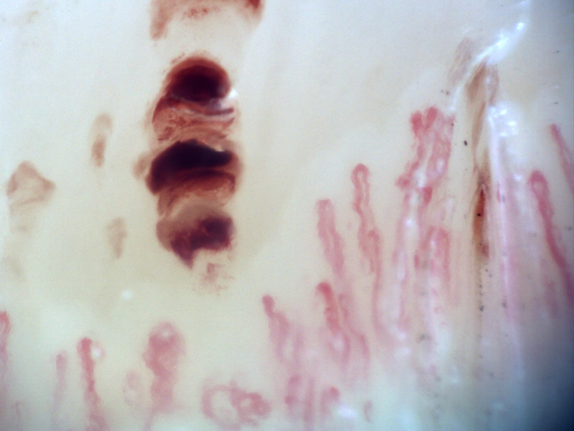
200x capillaroscopy image from systemic sclerosis showing a representative microvascular hemorrhagic lesion.
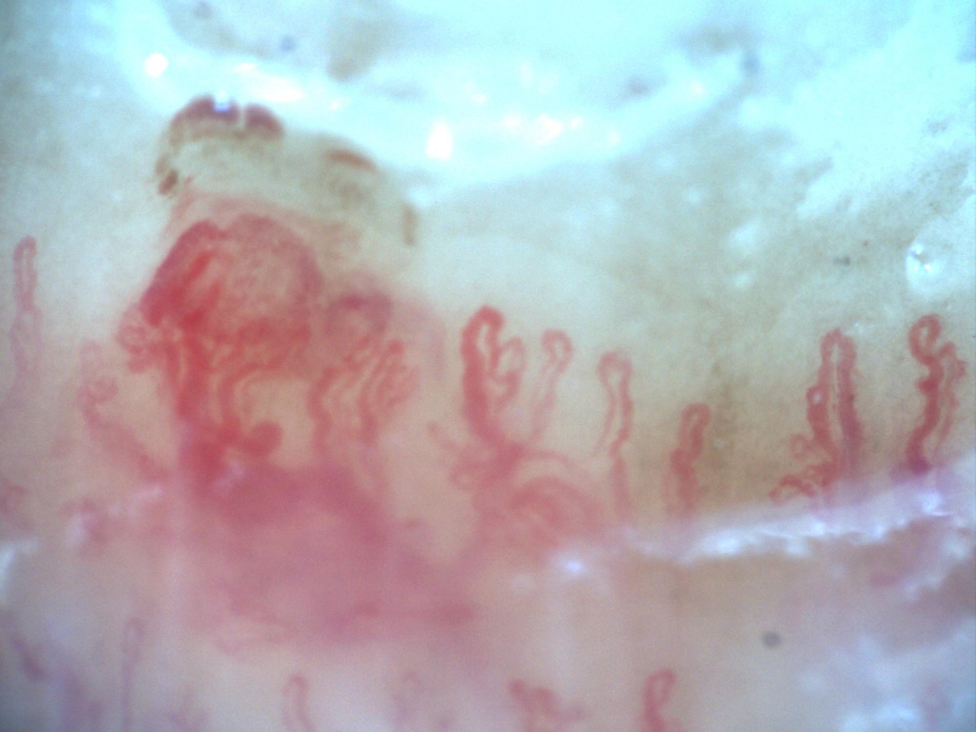
200x capillaroscopy image capturing a hemorrhage at the moment of its formation.
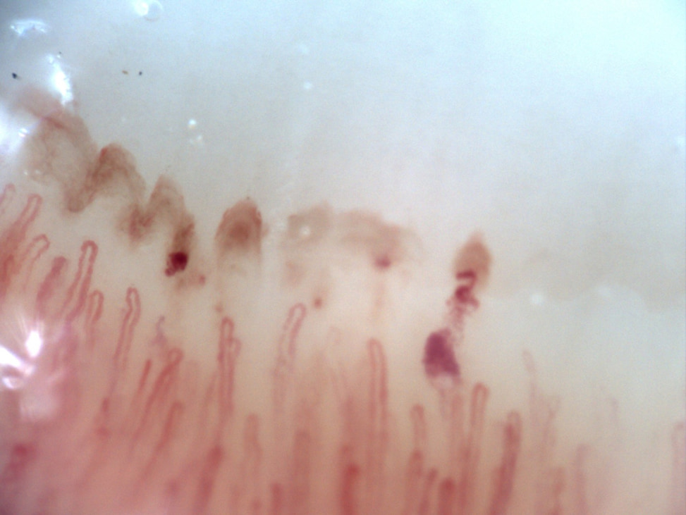 Preserved capillary density with anticoagulation-related hemorrhages
A 74-year-old male with no rheumatologic history, on apixaban for permanent atrial fibrillation. Nailfold video capillaroscopy of the right fourth finger demonstrates preserved capillary density without capillary dropout. There are no giants or enlarged capillaries. Hemorrhages are present, related to chronic anticoagulation.
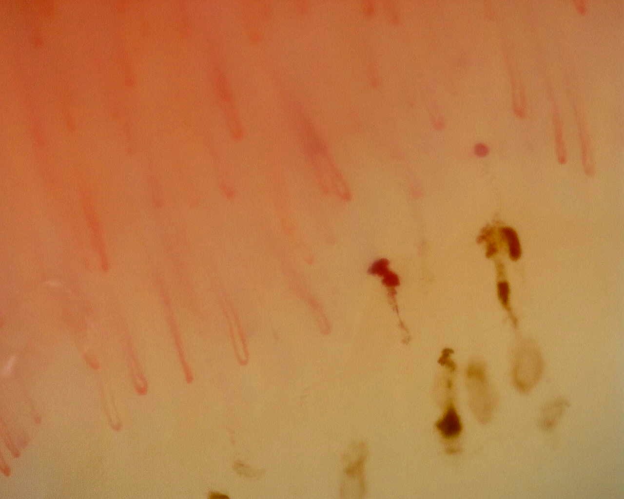 Nonspecific pattern: hemorrhagic field
Dark hemorrhagic deposits alongside capillary loops in the nonspecific reference study. Hemorrhage alone does not define a scleroderma pattern.
View the complete study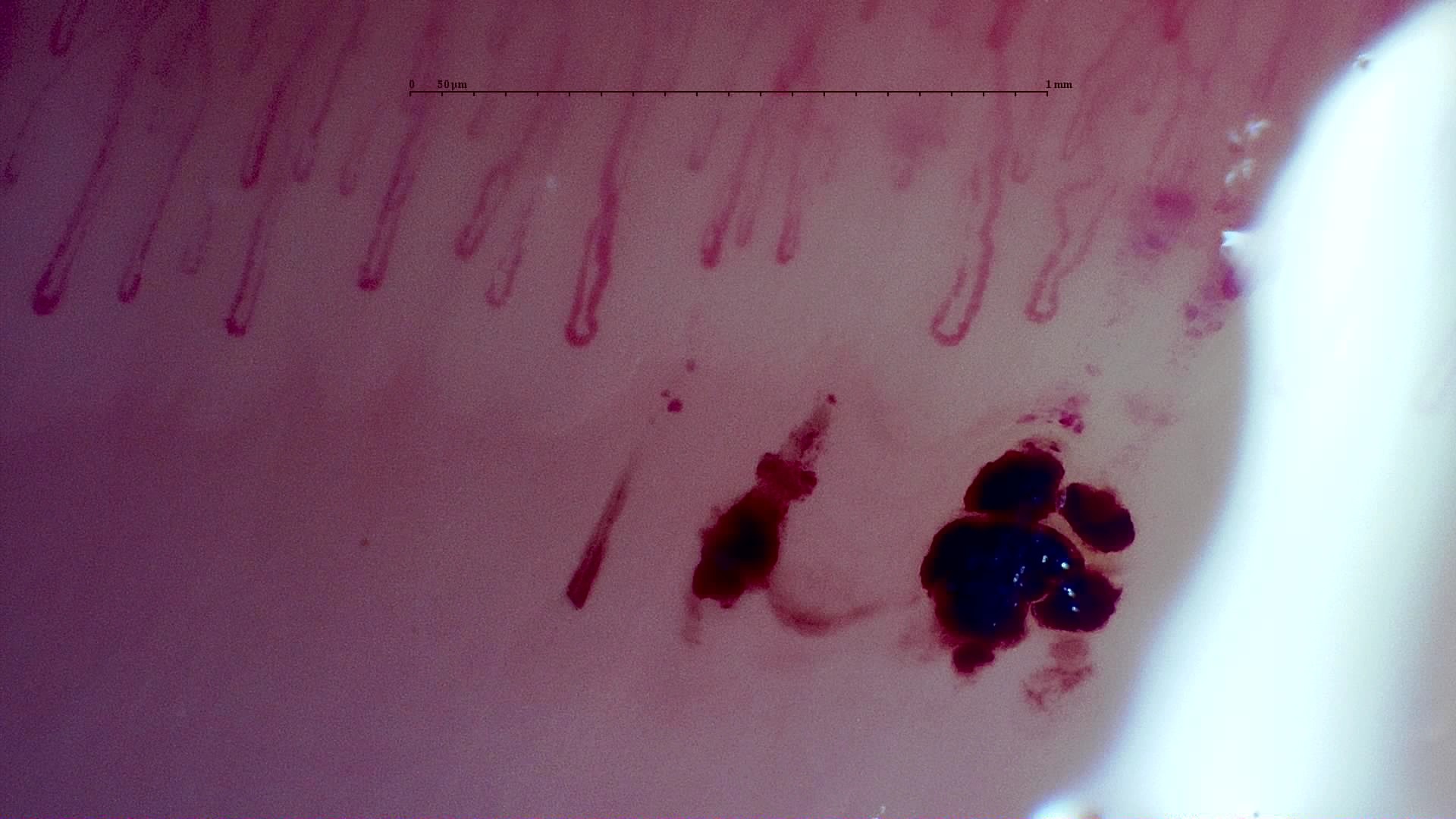 MPO-ANCA vasculitis: hemorrhagic deposits
Large dark hemorrhagic deposits below the capillary row in the published MPO-ANCA vasculitis case. Its complete study is described as a nonspecific pattern without giants.
View the complete study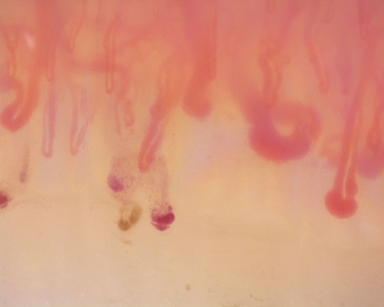 Anti-Scl-70 systemic sclerosis: enlarged loops and hemorrhages
Enlarged loops and dark hemorrhagic deposits in the anti-Scl-70-positive active-pattern case. The source report notes no avascular areas in the study.
View the complete studyComplete cases and studies
Non-Specific Pattern: Representative Case
The study shows preserved density, around 8.8 capillaries/mm, scattered dilated or tortuous loops, and hemorrhages around 0.46/mm, with no giant capillaries and no Cutolo SSc pattern.
Active SSc Cutolo Pattern: Representative Case
The study shows frequent giant capillaries and hemorrhages with reduced density and early architectural disorganization.
MPO-ANCA Vasculitis with Rapidly Progressive Glomerulonephritis
Capillaroscopy shows mildly reduced density (7.0 capillaries/mm), frequent capillary dilations, hemorrhages, and no giant capillaries, consistent with a non-specific microvascular pattern.
Diffuse Systemic Sclerosis (Anti-Scl-70+) with Active Cutolo Pattern
Capillaroscopy shows marked capillary reduction (5.86/mm), frequent dilations (54%), a high prevalence of giant capillaries (39.7%), and hemorrhages, without avascular areas, matching an active Cutolo pattern. The CSURI index has increased. Blood flow is homogeneous, with reduced linear velocity.
Juvenile Dermatomyositis with Gottron Lesions and a Scleroderma-Like Pattern
The baseline capillaroscopy is compatible with juvenile dermatomyositis and shows a severe scleroderma-like microangiopathy, with marked capillary loss, frequent enlarged and giant capillaries, hemorrhages, abnormal or arborizing shapes, and architectural disorganization.
Juvenile Dermatomyositis anti-NXP2 with Capillaroscopic Improvement
The June 2024 capillaroscopy showed an early scleroderma-like pattern compatible with juvenile dermatomyositis, including reduced density, frequent enlarged capillaries, sparse giant capillaries, and hemorrhages. The December 2024 follow-up showed disappearance of giant capillaries and evolution toward a non-specific pattern, with improved capillary density.
Mixed Connective Tissue Disease Presenting with Raynaud Phenomenon
The initial capillaroscopy showed an early scleroderma pattern, supporting the diagnosis in the appropriate clinical and serological context. On follow-up, clinical improvement was accompanied by improvement of capillaroscopic findings, with higher density, disappearance of giant capillaries, and evolution toward a non-specific pattern.
Anti-NXP2 Dermatomyositis with Capillaroscopic Improvement
The first study shows an active scleroderma/myositis-like microangiopathy with severe capillary loss, giant capillaries, arborizing ramifications, hemorrhages, architectural disorganization, and avascular areas. The follow-up study shows regression to a non-specific pattern with improved density and disappearance of giant capillaries and hemorrhages.
Systemic Sclerosis with anti-U3 RNP: Late Microangiopathy
There is marked capillary loss (4.9/mm), architectural disorganization, frequent dilations, giant capillaries in 14.9% of capillaries, and abnormal or ramified forms. This pattern was classified by the CAPI-Detect algorithm as active; however, due to the frequent features characteristic of late-stage involvement, it was ultimately classified as such in the final report.
References
- Smith V, et al. Standardisation of nailfold capillaroscopy for the assessment of patients with Raynaud’s phenomenon and systemic sclerosis. Autoimmun Rev. 2020;19:102458.
- Cutolo M, et al. Nailfold videocapillaroscopy assessment of microvascular damage in systemic sclerosis. J Rheumatol. 2000;27:155–160.
- Ingegnoli F, et al. Reporting items for capillaroscopy in clinical research on musculoskeletal diseases: a systematic review and international Delphi consensus. Rheumatology. 2021;60:1410–1418.
Scientific advisory board and contributors · Editorial policy