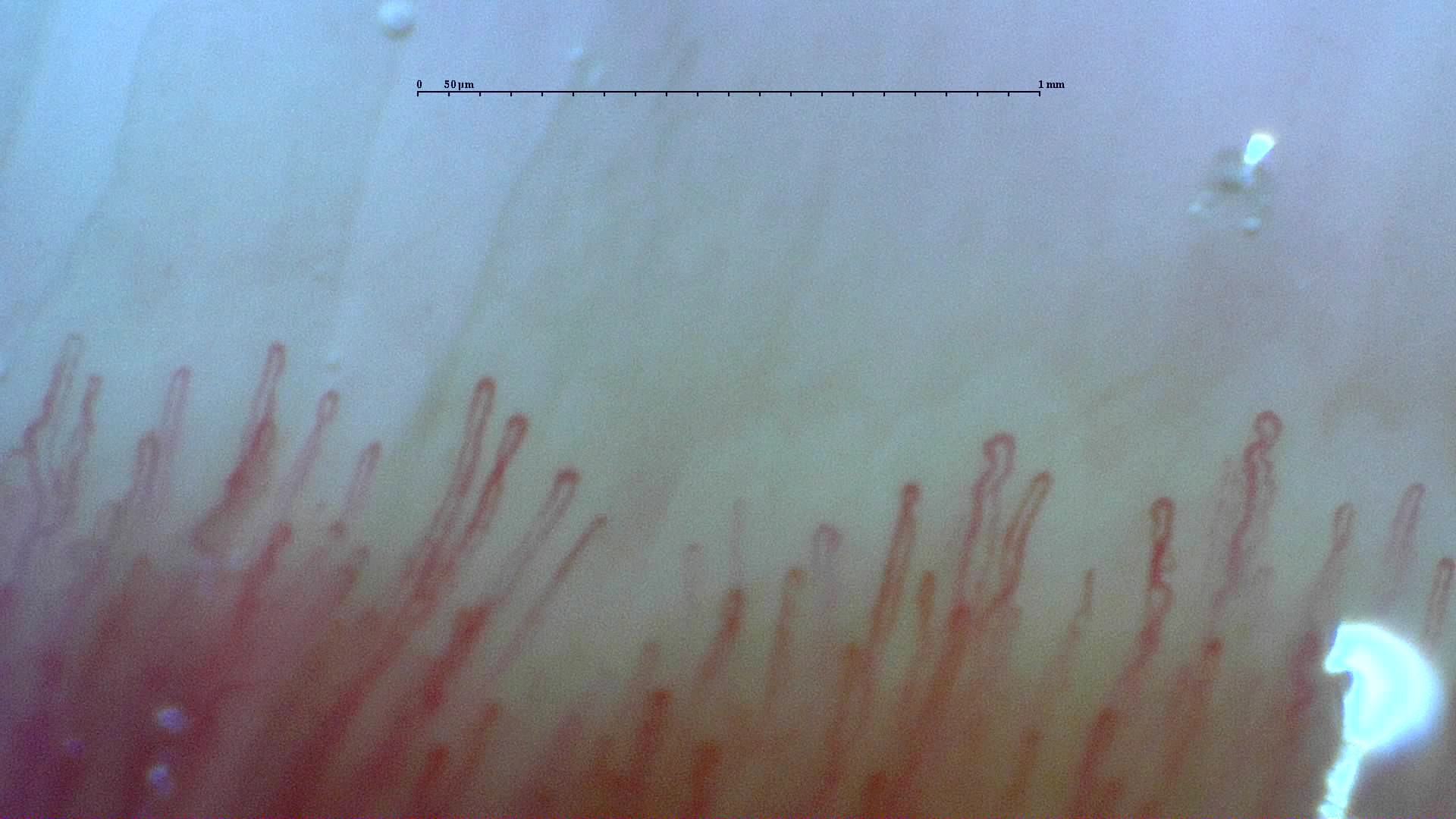
 Normal reference: distal capillary row
A field from the normal reference study, showing the distal row of narrow loops. Use the full study to compare density and shape across fingers.
View the complete studyHow to assess and measure
Count the distal-row capillaries over a calibrated length and divide by the length in millimeters. Use a consistent counting convention, avoid counting overlapping fields twice, and record which fingers and fields were assessable. Report the distribution as well as a study mean. Around 7 capillaries/mm is a commonly used lower reference in adults, but age, technique and image quality affect interpretation.
Interpretation and related diseases
Repeatedly reduced density supports capillary loss when acquisition is adequate. It is relevant in systemic sclerosis and inflammatory myopathy, but is not disease-specific. A focal avascular area and a low mean density describe related, different aspects of the study. Compare normal and abnormal fields and consider inter-digit variation before summarizing the examination.
Read the detailed clinical chapterCommon mistakes
- Counting deeper vessels as distal-row loops.
- Reporting capillaries per image without knowing the imaged length.
- Treating poor visibility, pressure collapse or an unassessable field as zero density.
- Applying an adult reference as a universal pediatric cutoff.
Real images and comparisons
Captions distinguish the illustrated finding from the study interpretation. Comparisons include variants, other clinical contexts or artifacts where available.
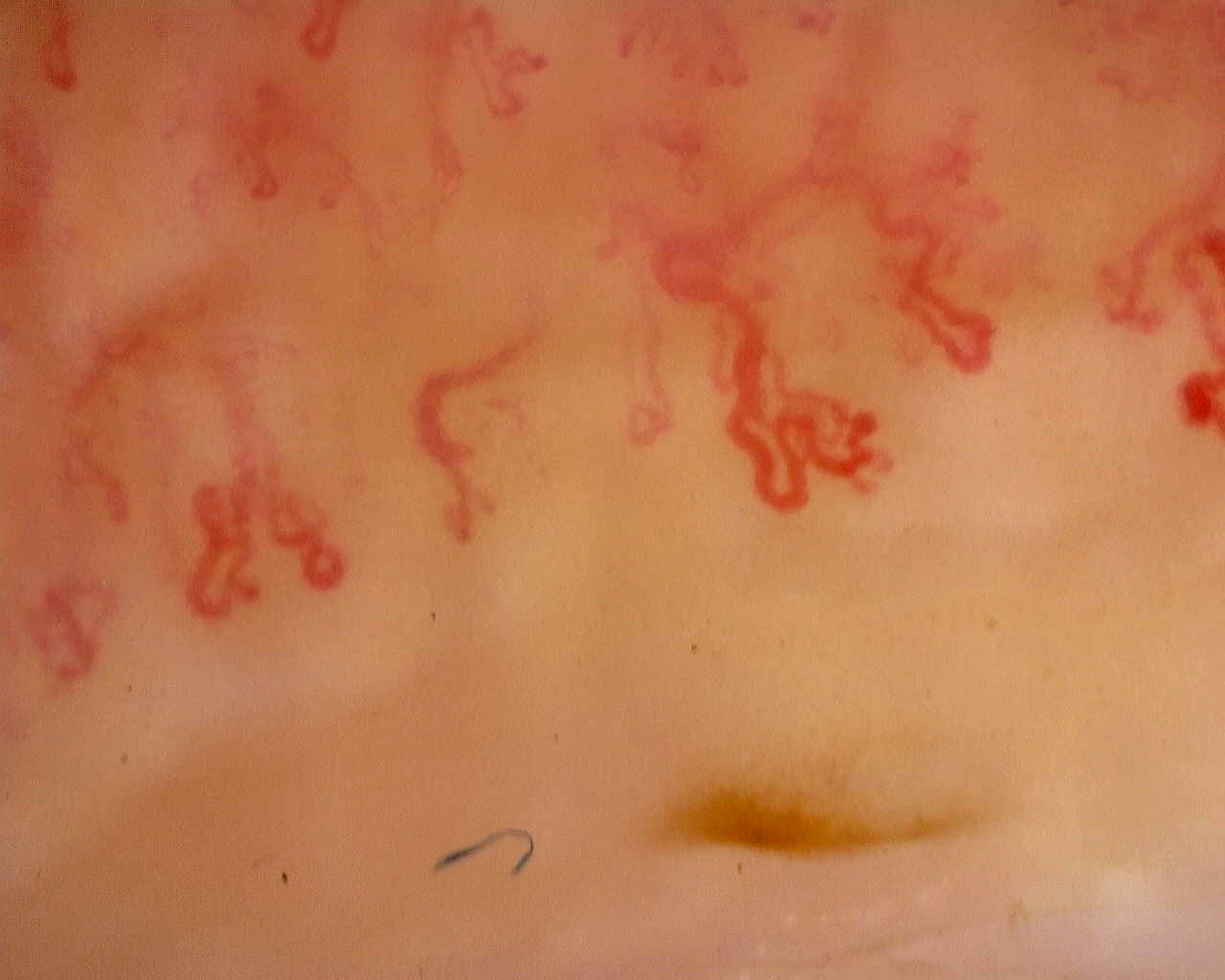 Late scleroderma pattern: disorganized loops
Irregular, ramified loops in a field from the late-pattern reference study. The full study provides context for capillary loss and distribution.
View the complete study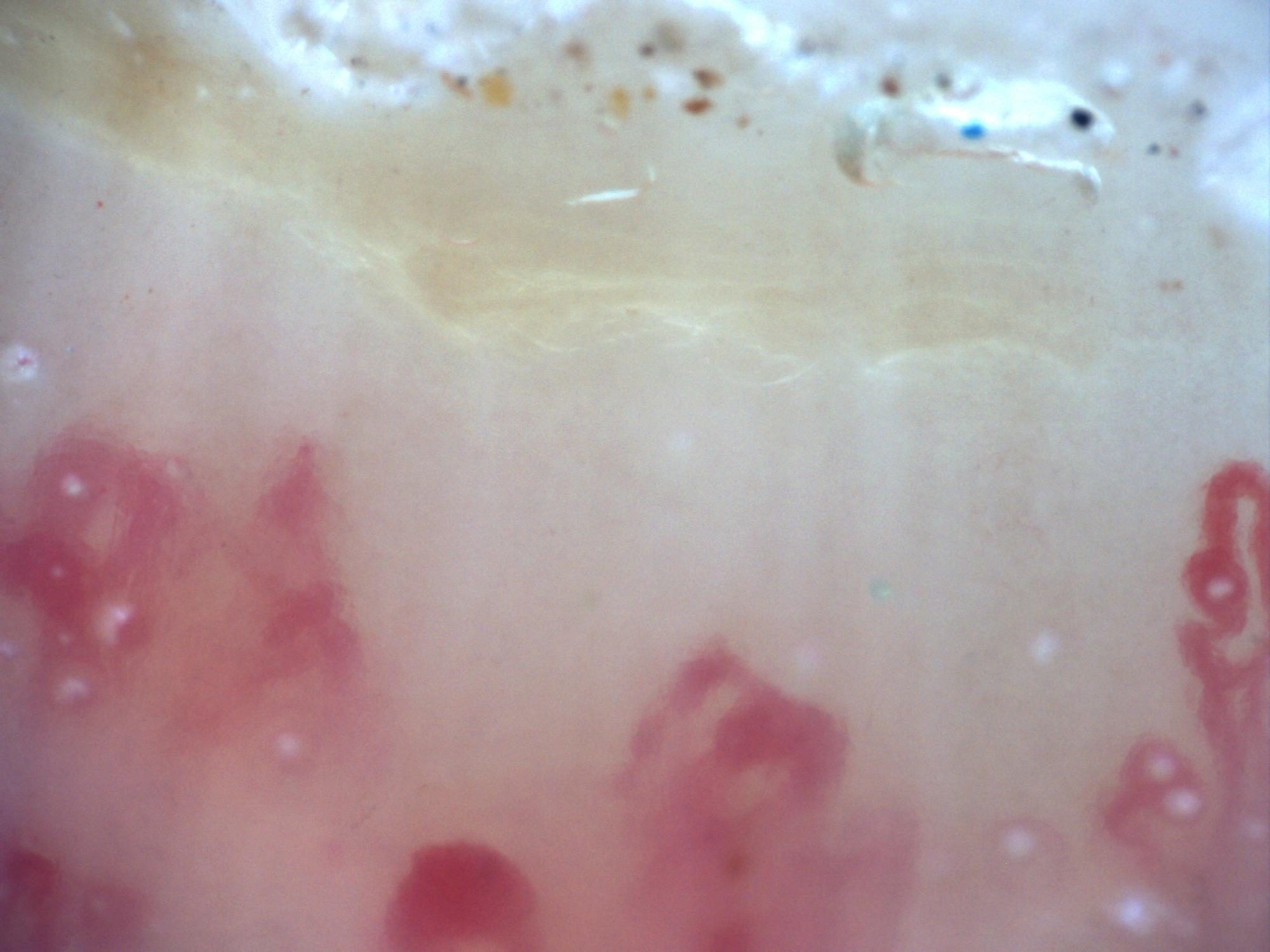
200x capillaroscopy image showing an extensive capillary-free space compatible with an avascular area.
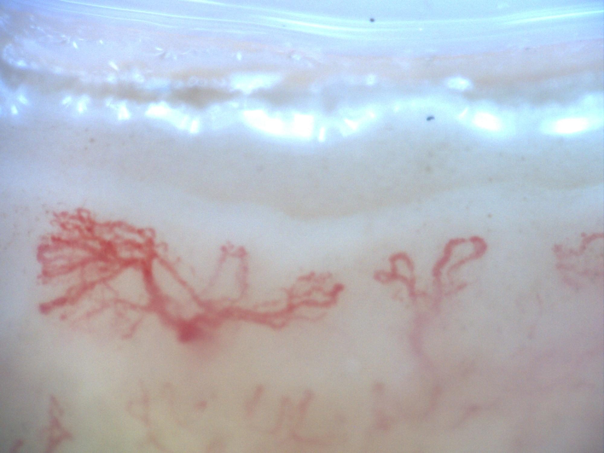
200x capillaroscopy image from a patient with systemic sclerosis showing a late pattern with marked disorganization and neoangiogenesis, without visible megacapillaries.
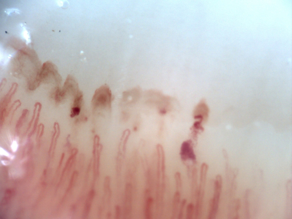 Preserved capillary density with anticoagulation-related hemorrhages
A 74-year-old male with no rheumatologic history, on apixaban for permanent atrial fibrillation. Nailfold video capillaroscopy of the right fourth finger demonstrates preserved capillary density without capillary dropout. There are no giants or enlarged capillaries. Hemorrhages are present, related to chronic anticoagulation.
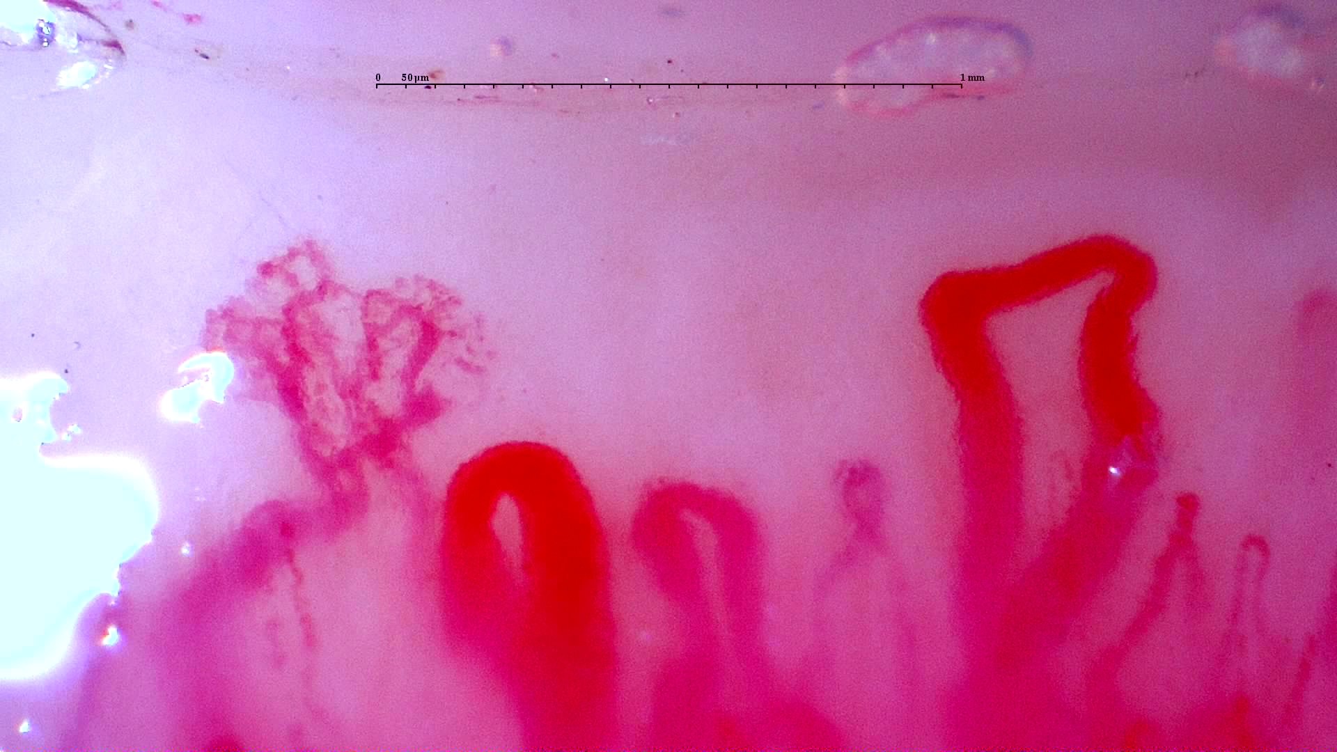 Anti-NXP2 dermatomyositis: initial enlarged loops
Prominent enlarged loops and a complex branching form in the initial anti-NXP2 dermatomyositis study. Compare this with the linked follow-up image and complete serial studies.
View the complete study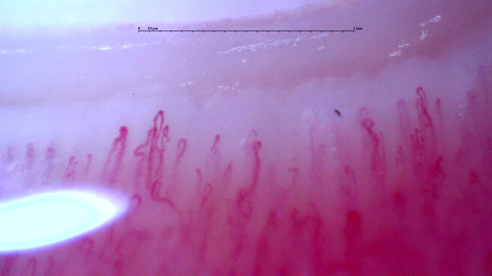 Anti-NXP2 dermatomyositis: follow-up field
Follow-up field from the adult anti-NXP2 dermatomyositis case. The source report documents improved study-wide density and disappearance of giants and hemorrhages; these are longitudinal study findings.
View the complete studyComplete cases and studies
Normal Pattern: Representative Case
The complete image set shows a regular distal row, preserved density, absent giant capillaries, and no avascular areas.
Late SSc Cutolo Pattern: Representative Case
The study shows severe capillary rarefaction, avascular areas, disorganization, and ramified or highly abnormal capillaries.
Anti-NXP2 Dermatomyositis with Capillaroscopic Improvement
The first study shows an active scleroderma/myositis-like microangiopathy with severe capillary loss, giant capillaries, arborizing ramifications, hemorrhages, architectural disorganization, and avascular areas. The follow-up study shows regression to a non-specific pattern with improved density and disappearance of giant capillaries and hemorrhages.
Anticentromere-Positive Systemic Sclerosis with Inter-Digit Pattern Heterogeneity
The study is notable for pronounced inter-digit and inter-field heterogeneity. L4C shows marked capillary loss (desertification), whereas L4A has relatively preserved, normal-appearing architecture. R2C and L3D show giant capillaries within a disorganized architecture. R4A demonstrates late-stage revascularization or neoangiogenesis—a morphology the contributor describes as a "post-late" pattern—while R5A shows transitional active-to-late features. The complete study is classified as a late scleroderma pattern, with a mean capillary density of 5.63/mm, enlarged capillaries in 43.5%, and giant capillaries in 2.5%. Image quality is variable; several fields are technically limited and should not be interpreted in isolation.
Juvenile Dermatomyositis with Gottron Lesions and a Scleroderma-Like Pattern
The baseline capillaroscopy is compatible with juvenile dermatomyositis and shows a severe scleroderma-like microangiopathy, with marked capillary loss, frequent enlarged and giant capillaries, hemorrhages, abnormal or arborizing shapes, and architectural disorganization.
Juvenile Dermatomyositis anti-NXP2 with Capillaroscopic Improvement
The June 2024 capillaroscopy showed an early scleroderma-like pattern compatible with juvenile dermatomyositis, including reduced density, frequent enlarged capillaries, sparse giant capillaries, and hemorrhages. The December 2024 follow-up showed disappearance of giant capillaries and evolution toward a non-specific pattern, with improved capillary density.
Juvenile Dermatomyositis anti-TIF1 with Active Scleroderma-Like Pattern
Capillaroscopy shows a severe active scleroderma-like pattern, with very low capillary density, numerous giant capillaries, frequent enlarged capillaries, arborizing abnormal shapes, and architectural disorganization. This morphology is highly suggestive of active microangiopathy in juvenile dermatomyositis.
Mixed Connective Tissue Disease Presenting with Raynaud Phenomenon
The initial capillaroscopy showed an early scleroderma pattern, supporting the diagnosis in the appropriate clinical and serological context. On follow-up, clinical improvement was accompanied by improvement of capillaroscopic findings, with higher density, disappearance of giant capillaries, and evolution toward a non-specific pattern.
Anti-TIF1γ Dermatomyositis with Scleroderma-Like Pattern
Capillaroscopy shows severely reduced capillary density (2.7/mm), architectural disorganization, frequent dilations, giant capillaries, and abnormal/ramified forms (2/3 on the semi-quantitative scale), consistent with a scleroderma-like pattern.
Systemic Sclerosis with anti-U3 RNP: Late Microangiopathy
There is marked capillary loss (4.9/mm), architectural disorganization, frequent dilations, giant capillaries in 14.9% of capillaries, and abnormal or ramified forms. This pattern was classified by the CAPI-Detect algorithm as active; however, due to the frequent features characteristic of late-stage involvement, it was ultimately classified as such in the final report.
References
- Smith V, et al. Standardisation of nailfold capillaroscopy for the assessment of patients with Raynaud’s phenomenon and systemic sclerosis. Autoimmun Rev. 2020;19:102458.
- Cutolo M, et al. Nailfold videocapillaroscopy assessment of microvascular damage in systemic sclerosis. J Rheumatol. 2000;27:155–160.
- Ingegnoli F, et al. Reporting items for capillaroscopy in clinical research on musculoskeletal diseases: a systematic review and international Delphi consensus. Rheumatology. 2021;60:1410–1418.
Scientific advisory board and contributors · Editorial policy