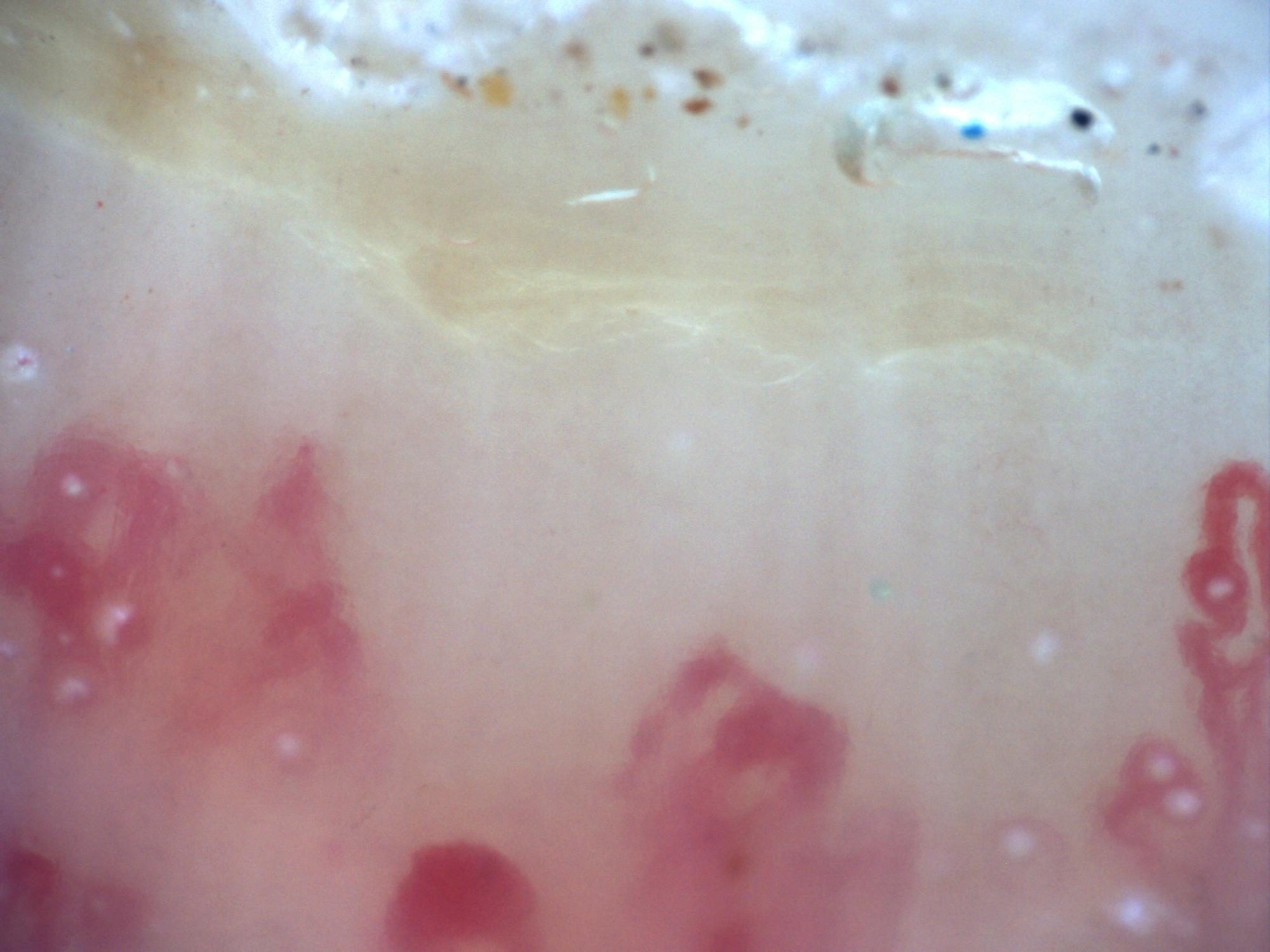
200x capillaroscopy image showing an extensive capillary-free space compatible with an avascular area.
How to assess and measure
Assess spacing in calibrated images and state the definition used by the reporting protocol. Describe the location, extent and distribution of gaps alongside density. Verify the same region with adequate focus, illumination and minimal probe pressure before labeling capillary dropout. Do not attach a micrometer width to an uncalibrated screenshot.
Interpretation and related diseases
Avascular areas, severe capillary loss, disorganization and ramified loops are characteristic components of late scleroderma-pattern damage. They may also occur in severe myositis-associated microangiopathy. The pressure-artifact comparison below illustrates an important mimic: capillaries can become invisible without having been structurally lost.
Read the detailed clinical chapterCommon mistakes
- Calling an out-of-focus, reflective or compressed region avascular.
- Inferring whole-study density from a single gap.
- Assuming that every active scleroderma-pattern study must contain avascular areas.
Real images and comparisons
Captions distinguish the illustrated finding from the study interpretation. Comparisons include variants, other clinical contexts or artifacts where available.
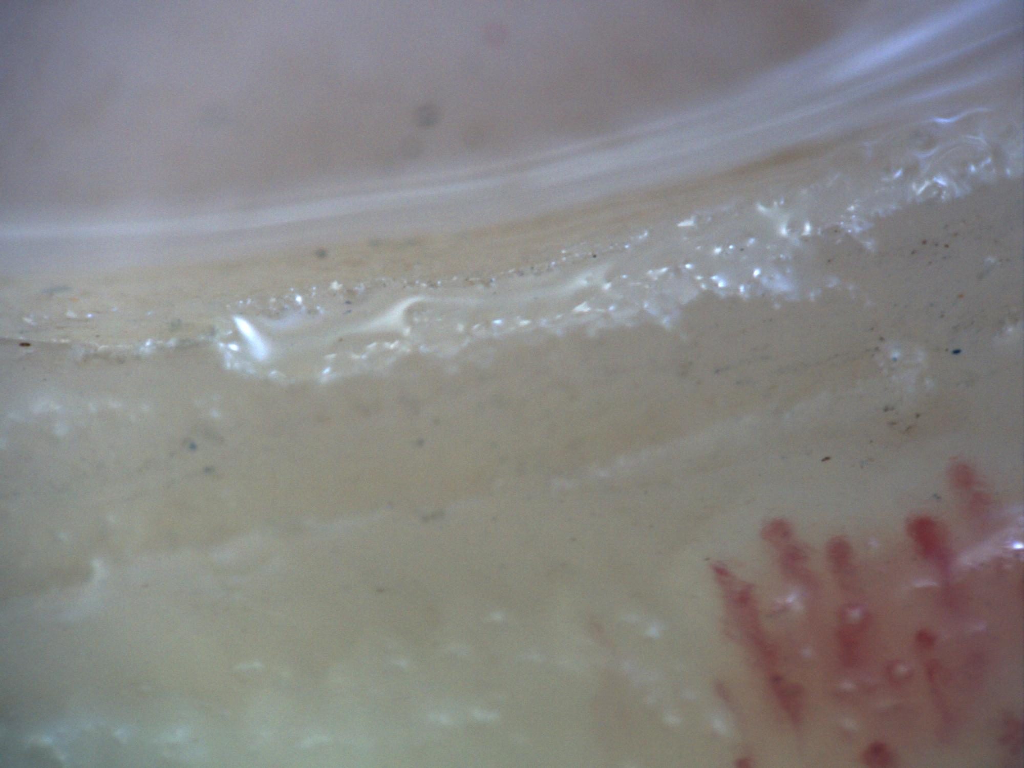
200x capillaroscopy image showing excessive pressure on the nail, with disappearance of the visible capillary bed along the left margin.
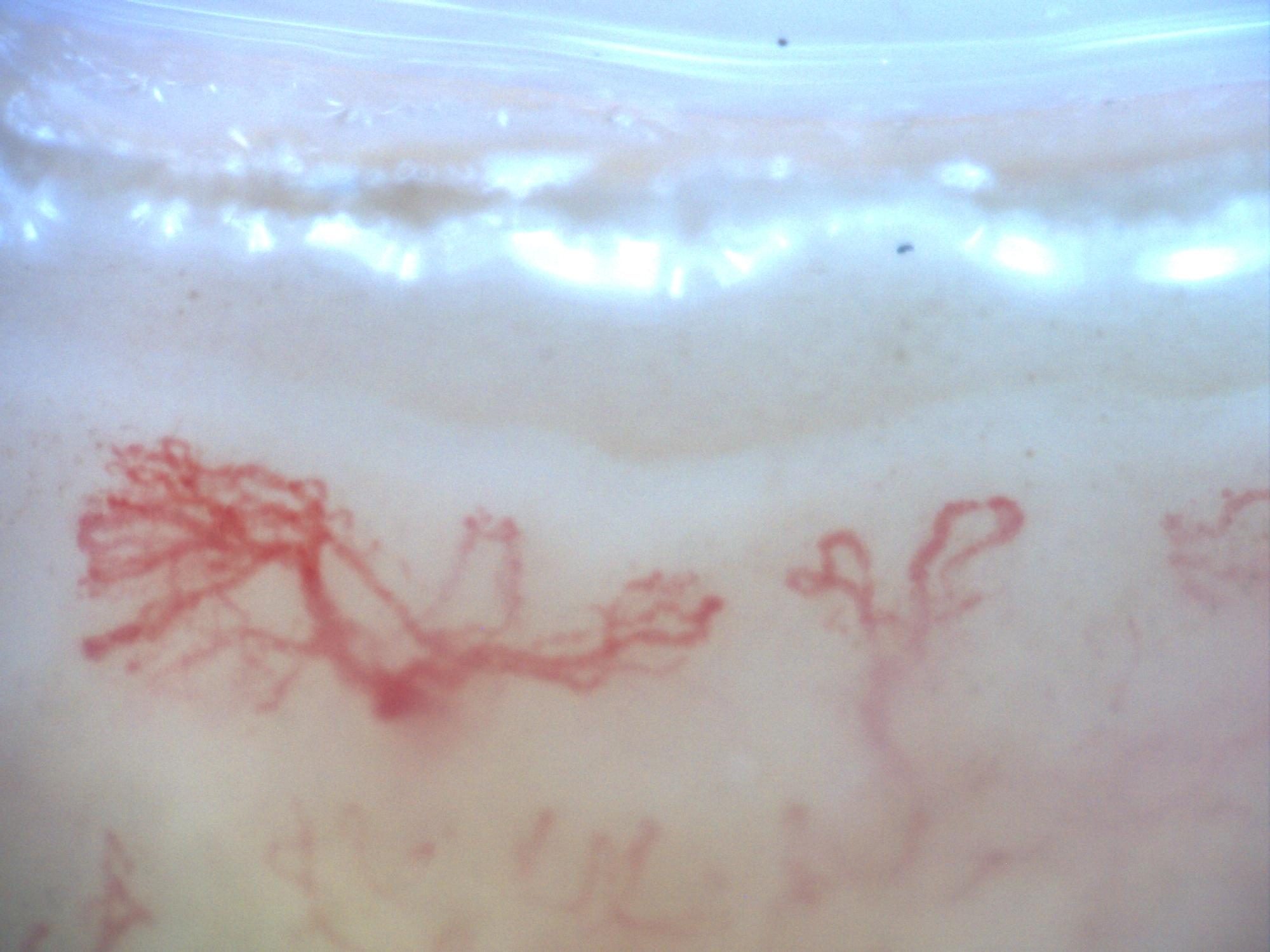
200x capillaroscopy image from a patient with systemic sclerosis showing a late pattern with marked disorganization and neoangiogenesis, without visible megacapillaries.
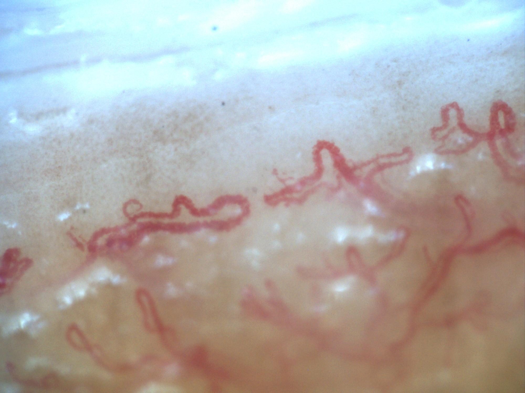
Second 200x capillaroscopy image from systemic sclerosis with a late pattern, marked disorganization, and neoangiogenesis.
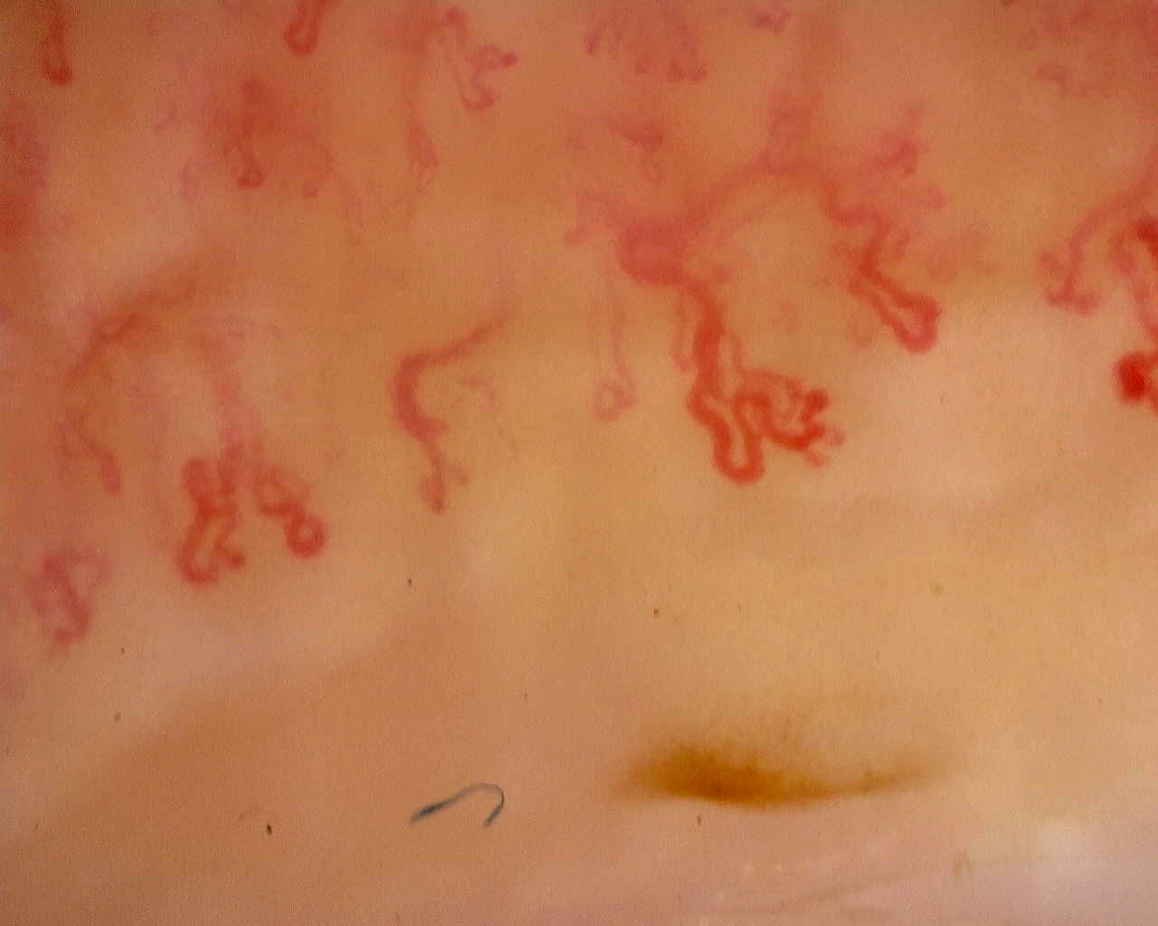 Late scleroderma pattern: disorganized loops
Irregular, ramified loops in a field from the late-pattern reference study. The full study provides context for capillary loss and distribution.
View the complete studyComplete cases and studies
Late SSc Cutolo Pattern: Representative Case
The study shows severe capillary rarefaction, avascular areas, disorganization, and ramified or highly abnormal capillaries.
Anticentromere-Positive Systemic Sclerosis with Inter-Digit Pattern Heterogeneity
The study is notable for pronounced inter-digit and inter-field heterogeneity. L4C shows marked capillary loss (desertification), whereas L4A has relatively preserved, normal-appearing architecture. R2C and L3D show giant capillaries within a disorganized architecture. R4A demonstrates late-stage revascularization or neoangiogenesis—a morphology the contributor describes as a "post-late" pattern—while R5A shows transitional active-to-late features. The complete study is classified as a late scleroderma pattern, with a mean capillary density of 5.63/mm, enlarged capillaries in 43.5%, and giant capillaries in 2.5%. Image quality is variable; several fields are technically limited and should not be interpreted in isolation.
Anti-NXP2 Dermatomyositis with Capillaroscopic Improvement
The first study shows an active scleroderma/myositis-like microangiopathy with severe capillary loss, giant capillaries, arborizing ramifications, hemorrhages, architectural disorganization, and avascular areas. The follow-up study shows regression to a non-specific pattern with improved density and disappearance of giant capillaries and hemorrhages.
References
- Smith V, et al. Standardisation of nailfold capillaroscopy for the assessment of patients with Raynaud’s phenomenon and systemic sclerosis. Autoimmun Rev. 2020;19:102458.
- Cutolo M, et al. Nailfold videocapillaroscopy assessment of microvascular damage in systemic sclerosis. J Rheumatol. 2000;27:155–160.
- Ingegnoli F, et al. Reporting items for capillaroscopy in clinical research on musculoskeletal diseases: a systematic review and international Delphi consensus. Rheumatology. 2021;60:1410–1418.
Scientific advisory board and contributors · Editorial policy