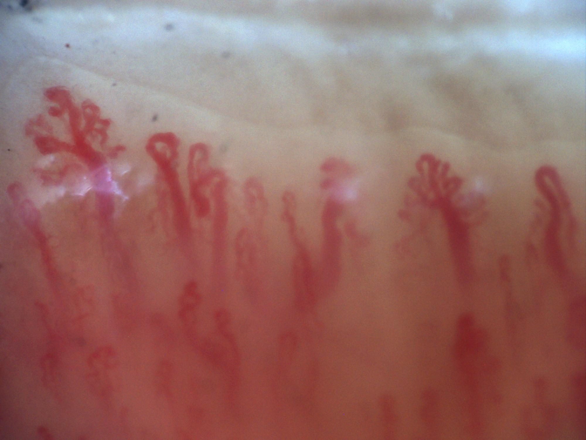
200x capillaroscopy image with a branching capillary on the left side of the field.
How to assess and measure
Assess morphology in focused, non-overlapping fields. Distinguish true branches from separate loops crossing in projection. When reporting a proportion of abnormal shapes, state the classification and denominator; count according to one protocol. Record accompanying density loss and architectural disorganization rather than reducing the study to one shape score.
Interpretation and related diseases
Ramifications and disorganization are prominent in late scleroderma-pattern microangiopathy and can also be seen in dermatomyositis and mixed connective tissue disease. The term neoangiogenesis describes the interpretation of a remodeling pattern; a single unusual loop does not directly demonstrate new-vessel formation over time.
Read the detailed clinical chapterCommon mistakes
- Equating simple tortuosity with complex branching.
- Assuming bushy capillaries are specific to systemic sclerosis.
- Using an algorithmic shape category without checking the image and overall pattern.
Real images and comparisons
Captions distinguish the illustrated finding from the study interpretation. Comparisons include variants, other clinical contexts or artifacts where available.
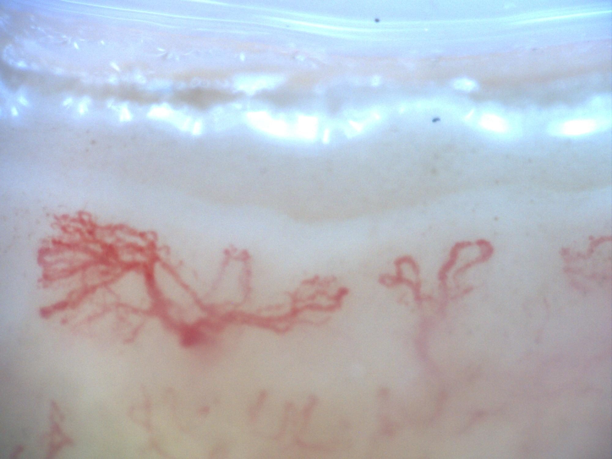
200x capillaroscopy image from a patient with systemic sclerosis showing a late pattern with marked disorganization and neoangiogenesis, without visible megacapillaries.
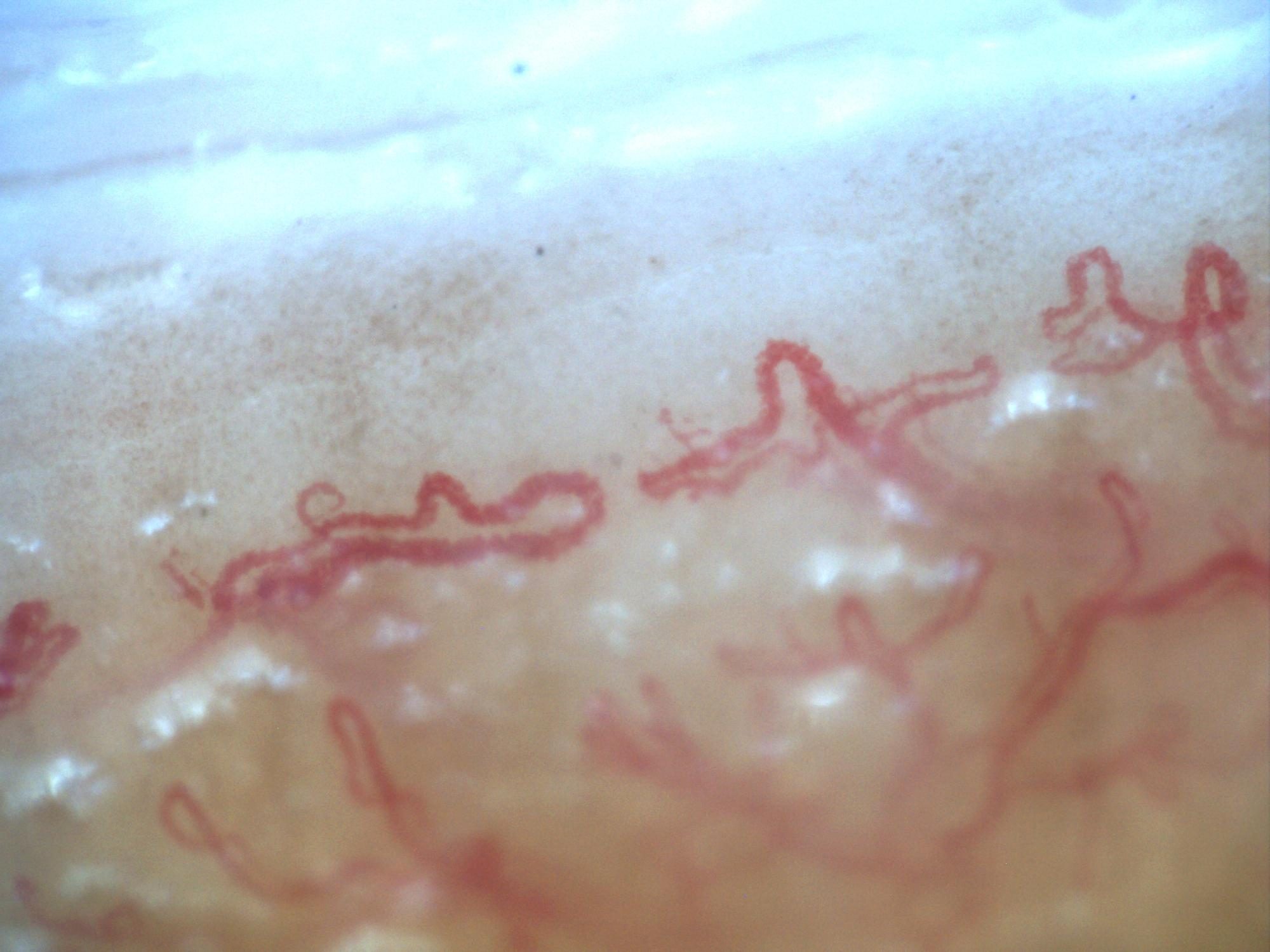
Second 200x capillaroscopy image from systemic sclerosis with a late pattern, marked disorganization, and neoangiogenesis.
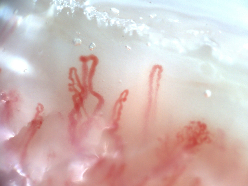 Dermatomyositis with giant and ramified capillaries
25-year-old male with anti-TIF-1γ antibody-positive dermatomyositis. Nailfold video capillaroscopy demonstrates significant microvascular architectural abnormalities characterized by enlarged and giant capillaries and multiple ramified (bushy) capillaries consistent with neoangiogenesis. No microhemorrhages are identified.
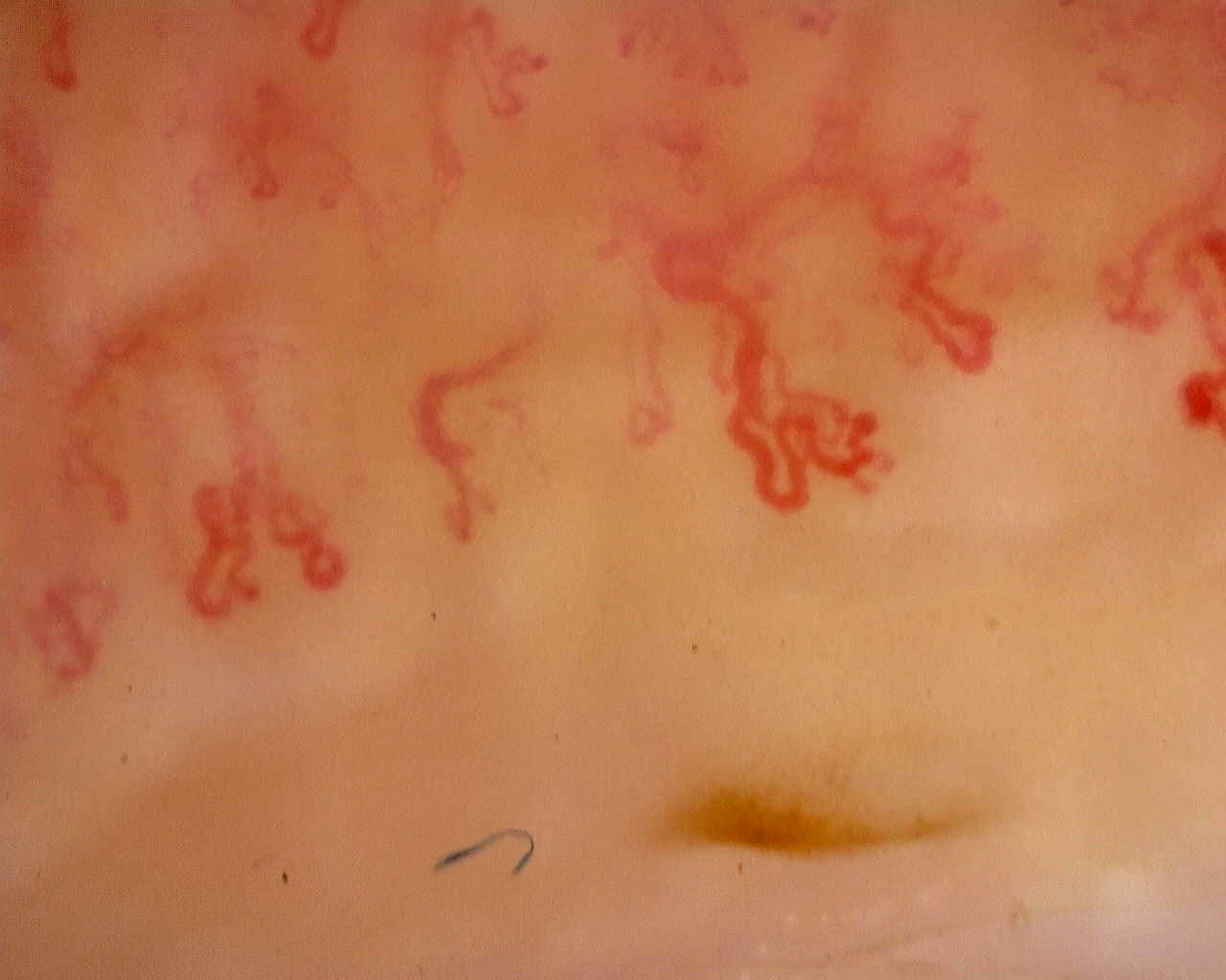 Late scleroderma pattern: disorganized loops
Irregular, ramified loops in a field from the late-pattern reference study. The full study provides context for capillary loss and distribution.
View the complete study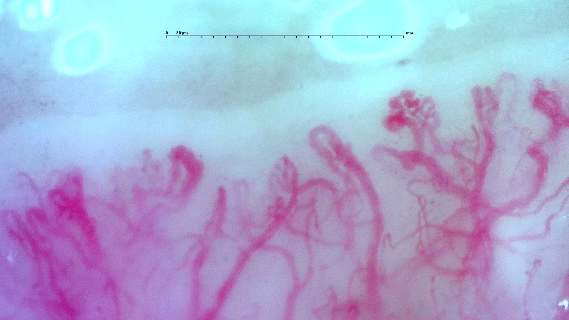 Anti-TIF1γ dermatomyositis: complex vascular shapes
Complex, winding and ramified vascular forms in the anti-TIF1γ dermatomyositis case. A scleroderma-like image appearance does not establish systemic sclerosis.
View the complete study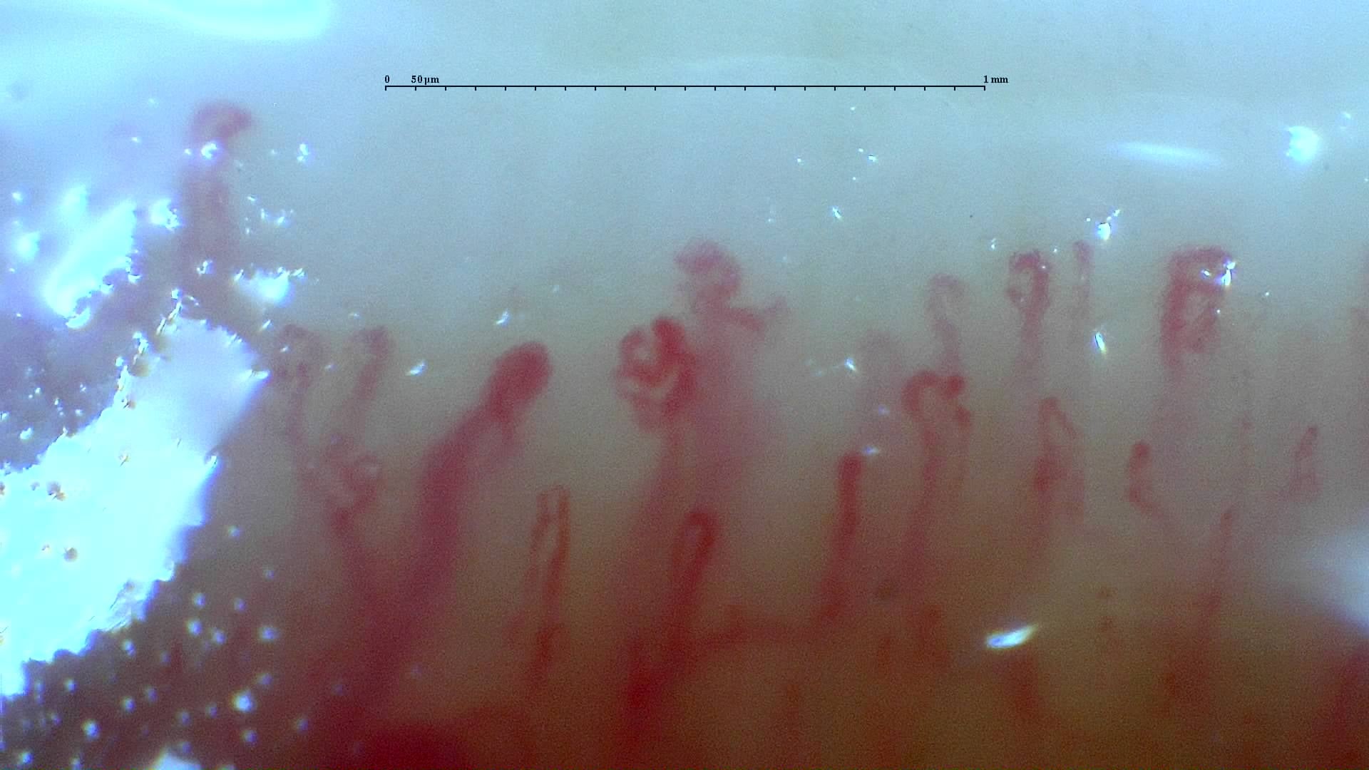 Mixed connective tissue disease: irregular branching forms
Irregular and branching forms in the anti-RNP-positive mixed connective tissue disease case. The source report describes a scleroderma-like pattern without an assignable Cutolo subtype.
View the complete studyComplete cases and studies
Late SSc Cutolo Pattern: Representative Case
The study shows severe capillary rarefaction, avascular areas, disorganization, and ramified or highly abnormal capillaries.
Anti-Mi2β Dermatomyositis in Active Disease
There is marked capillary loss (4.8/mm), architectural disorganization, frequent dilations, giant capillaries, and abnormal/branched shapes, indicating severe active microangiopathy.
Mixed Connective Tissue Disease with an Unclassifiable Scleroderma-Like Pattern
Capillaroscopy shows megacapillaries, but their distribution does not fit the typical organization of any early, active, or late Cutolo subtype. The overall morphology is therefore classified as a scleroderma-like pattern. Arborizing capillaries are also evident. The quantitative dataset reports a mean capillary density of 5.69/mm, enlarged capillaries in 78.7%, megacapillaries in 6.0%, and ramified forms in 4.7%.
Anti-TIF1γ Dermatomyositis with Scleroderma-Like Pattern
Capillaroscopy shows severely reduced capillary density (2.7/mm), architectural disorganization, frequent dilations, giant capillaries, and abnormal/ramified forms (2/3 on the semi-quantitative scale), consistent with a scleroderma-like pattern.
Juvenile Dermatomyositis with Gottron Lesions and a Scleroderma-Like Pattern
The baseline capillaroscopy is compatible with juvenile dermatomyositis and shows a severe scleroderma-like microangiopathy, with marked capillary loss, frequent enlarged and giant capillaries, hemorrhages, abnormal or arborizing shapes, and architectural disorganization.
Juvenile Dermatomyositis anti-TIF1 with Active Scleroderma-Like Pattern
Capillaroscopy shows a severe active scleroderma-like pattern, with very low capillary density, numerous giant capillaries, frequent enlarged capillaries, arborizing abnormal shapes, and architectural disorganization. This morphology is highly suggestive of active microangiopathy in juvenile dermatomyositis.
Anti-NXP2 Dermatomyositis with Capillaroscopic Improvement
The first study shows an active scleroderma/myositis-like microangiopathy with severe capillary loss, giant capillaries, arborizing ramifications, hemorrhages, architectural disorganization, and avascular areas. The follow-up study shows regression to a non-specific pattern with improved density and disappearance of giant capillaries and hemorrhages.
Systemic Sclerosis with anti-U3 RNP: Late Microangiopathy
There is marked capillary loss (4.9/mm), architectural disorganization, frequent dilations, giant capillaries in 14.9% of capillaries, and abnormal or ramified forms. This pattern was classified by the CAPI-Detect algorithm as active; however, due to the frequent features characteristic of late-stage involvement, it was ultimately classified as such in the final report.
References
- Smith V, et al. Standardisation of nailfold capillaroscopy for the assessment of patients with Raynaud’s phenomenon and systemic sclerosis. Autoimmun Rev. 2020;19:102458.
- Cutolo M, et al. Nailfold videocapillaroscopy assessment of microvascular damage in systemic sclerosis. J Rheumatol. 2000;27:155–160.
- Ingegnoli F, et al. Reporting items for capillaroscopy in clinical research on musculoskeletal diseases: a systematic review and international Delphi consensus. Rheumatology. 2021;60:1410–1418.
Scientific advisory board and contributors · Editorial policy