Clinical Cases
Real-world case studies with capillaroscopic images, interpretation, and clinical correlation.
Case-Based Learning
Clinical Cases
Clinical cases demonstrate practical application of capillaroscopic interpretation in real-world scenarios. Each case card includes diagnosis-focused clinical context, structured capillaroscopy interpretation, and key findings designed for rapid review and searchability.
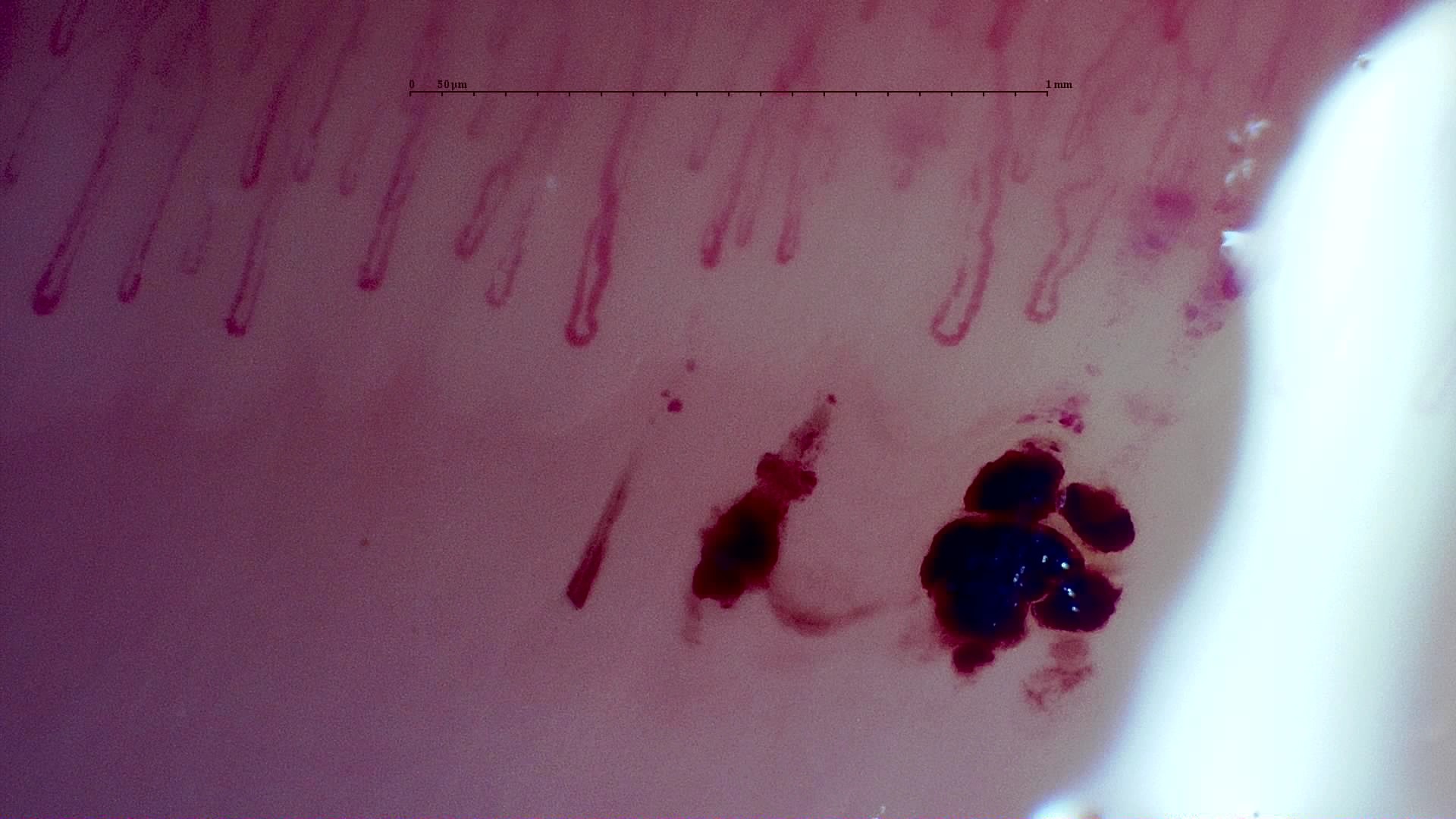 MPO-ANCA Vasculitis with Rapidly Progressive GlomerulonephritisANCA-Associated Vasculitis (MPO+)
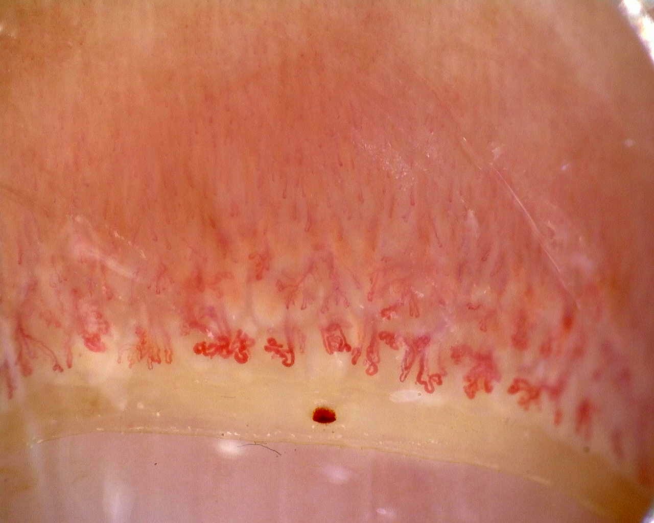 Anti-Mi2β Dermatomyositis in Active DiseaseDermatomyositis (anti-Mi2β+)
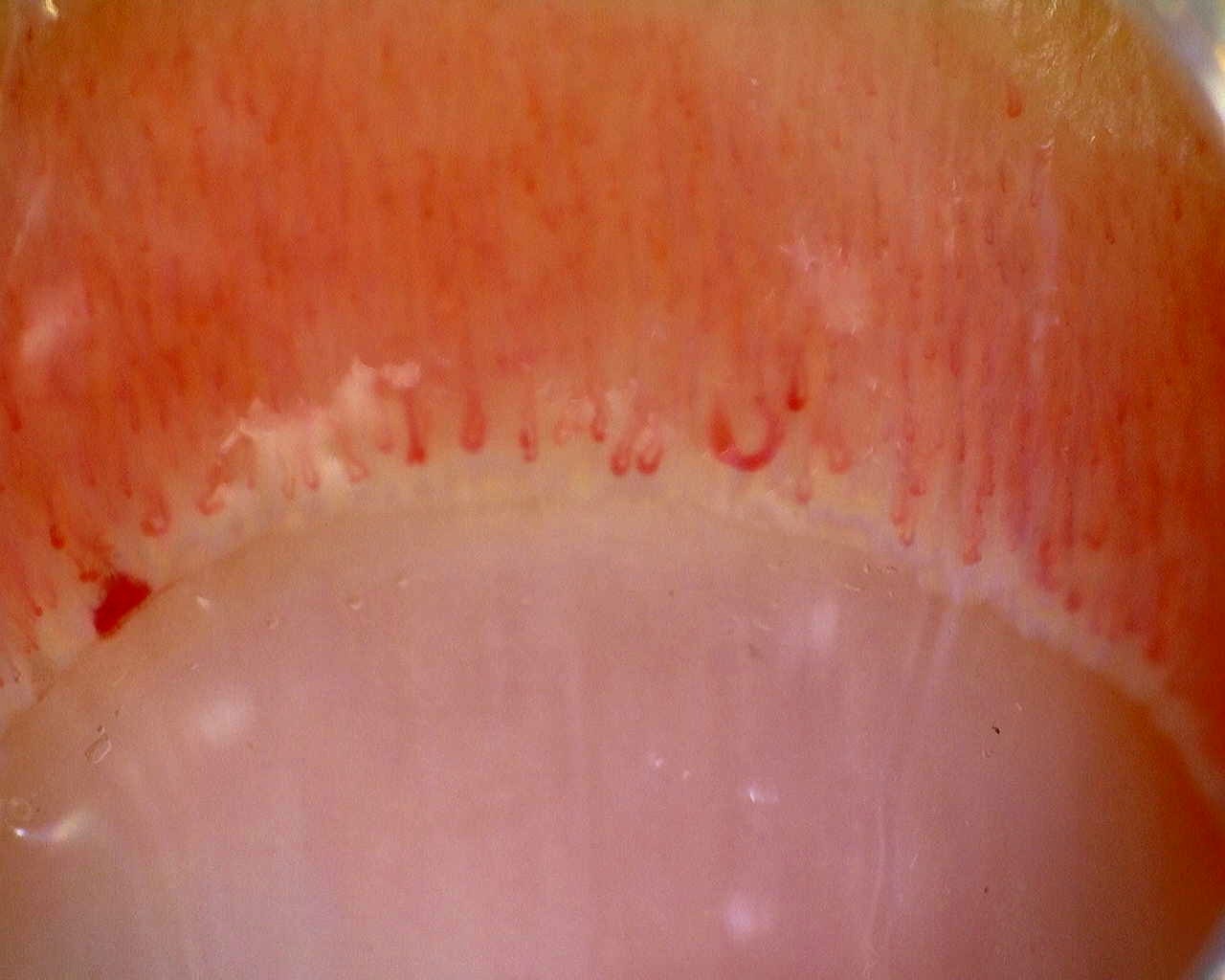 Limited Cutaneous Systemic Sclerosis (ACA+) with Active Cutolo PatternLimited Cutaneous Systemic Sclerosis (ACA+)
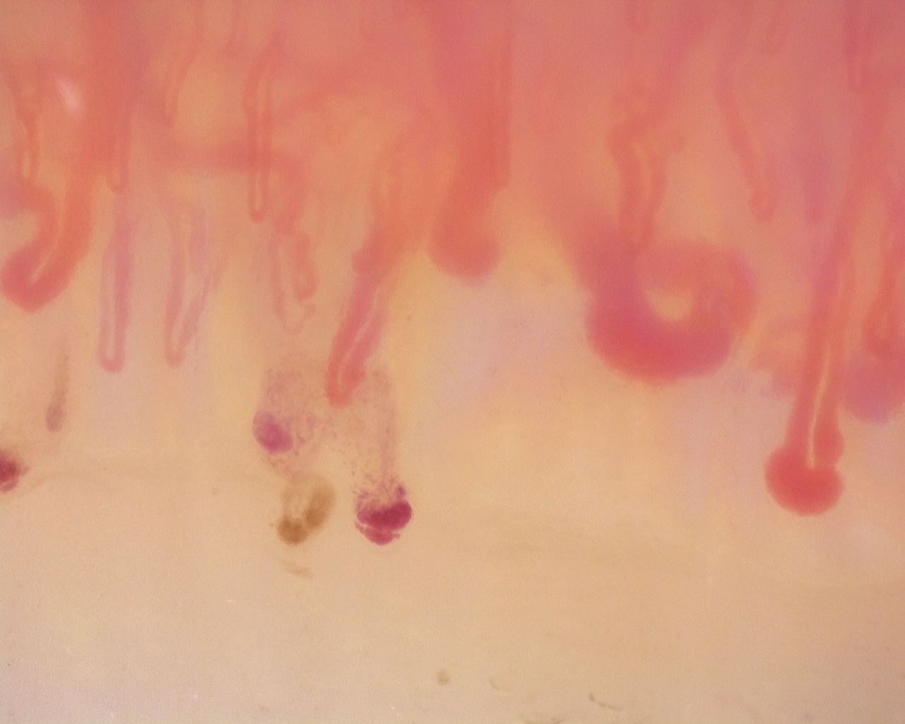 Diffuse Systemic Sclerosis (Anti-Scl-70+) with Active Cutolo PatternDiffuse Systemic Sclerosis (anti-Scl-70+)
Click tags to filter cases. Click again to remove a tag.
Pathology
Severity
Pattern
Showing 13 of 13 cases
Active filters
MPO-ANCA Vasculitis with Rapidly Progressive Glomerulonephritis
A 57-year-old man with newly diagnosed MPO-ANCA vasculitis and rapidly progressive glomerulonephritis.
Clinical context: The patient presented with renal failure and nephritic syndrome, and kidney biopsy confirmed rapidly progressive glomerulonephritis in the setting of MPO-ANCA vasculitis.
Capillaroscopy: Capillaroscopy shows mildly reduced density (7.0 capillaries/mm), frequent capillary dilations, hemorrhages, and no giant capillaries, consistent with a non-specific microvascular pattern.
Key Capillaroscopic Findings:
- • Mild capillary density reduction (7.0/mm)
- • Frequent capillary dilations (58.2%)
- • Hemorrhages (0.5/mm), without giant capillaries
Anti-Mi2β Dermatomyositis in Active Disease
A 32-year-old woman with active anti-Mi2β-positive dermatomyositis and marked microvascular damage on capillaroscopy.
Clinical context: After 3 months of progressive symptoms, diagnosis was established based on proximal muscle weakness, Gottron papules, heliotrope rash, and hyperCKemia (~2500).
Capillaroscopy: There is marked capillary loss (4.8/mm), architectural disorganization, frequent dilations, giant capillaries, and abnormal/branched shapes, indicating severe active microangiopathy.
Key Capillaroscopic Findings:
- • Severe capillary loss (4.8/mm)
- • Giant capillaries (8.8%) and frequent dilations (59.4%)
- • Abnormal/branched capillaries (32.1%) with vascular remodeling
Limited Cutaneous Systemic Sclerosis (ACA+) with Active Cutolo Pattern
A 38-year-old woman with limited cutaneous systemic sclerosis, anti-centromere antibodies, and established active microangiopathy.
Clinical context: This case includes a 5-year history of Raynaud's phenomenon, esophageal dysmotility, and interstitial lung disease with an NSIP pattern, without current indirect echocardiographic signs of pulmonary hypertension.
Capillaroscopy: Capillaroscopy shows marked capillary loss (5.3/mm), frequent dilations, high prevalence of giant capillaries (27.5%), and hemorrhages, matching an active Cutolo pattern.
Key Capillaroscopic Findings:
- • Marked capillary loss (5.3/mm)
- • High frequency of giant capillaries (27.5%)
- • Frequent capillary dilations (61.6%) with hemorrhages
Diffuse Systemic Sclerosis (Anti-Scl-70+) with Active Cutolo Pattern
A 19-year-old man with diffuse systemic sclerosis, positive anti-Scl-70 antibodies, and established active microangiopathy.
Clinical context: The case includes a 10-year history of acrocyanosis with tingling and burning sensations, more pronounced in cold environments. He has mild esophageal dysmotility and several apical subpleural fibrous nodules on chest CT, without current indirect echocardiographic signs of pulmonary hypertension. He is currently being treated with mycophenolate mofetil and amlodipine.
Capillaroscopy: Capillaroscopy shows marked capillary reduction (5.86/mm), frequent dilations (54%), a high prevalence of giant capillaries (39.7%), and hemorrhages, without avascular areas, matching an active Cutolo pattern. The CSURI index has increased. Blood flow is homogeneous, with reduced linear velocity.
Key Capillaroscopic Findings:
- • Marked capillary loss (5.86/mm)
- • High frequency of giant capillaries (39.7%)
- • Frequent capillary dilations (54%)
- • Hemorrhages indicating persistent vascular damage
- • No avascular areas
Systemic Sclerosis with anti-U3 RNP: Late Microangiopathy
Patient with systemic sclerosis, anti-U3 RNP antibodies, and severe microvascular disease with features of the late Cutolo pattern.
Clinical context: Patient under follow-up by internal medicine, in whom a positive ANA was documented, with findings compatible with nonspecific interstitial pneumonia. The patient had an ANA titer greater than 1:1280 with an AC-9 granular pattern. Subsequent studies showed high-titer positivity for anti-U3 RNP antibodies. The patient presented with salt-and-pepper pigmentation, generalized cutaneous sclerosis, and sclerodactyly.
Capillaroscopy: There is marked capillary loss (4.9/mm), architectural disorganization, frequent dilations, giant capillaries in 14.9% of capillaries, and abnormal or ramified forms. This pattern was classified by the CAPI-Detect algorithm as active; however, due to the frequent features characteristic of late-stage involvement, it was ultimately classified as such in the final report.
Key Capillaroscopic Findings:
- • Marked capillary loss (4.9/mm)
- • Giant capillaries (14.9%)
- • Architectural disorganization
- • Hemorrhages indicating persistent vascular damage
Anticentromere-Positive Systemic Sclerosis with Inter-Digit Pattern Heterogeneity
A 61-year-old Latin American woman with long-standing Raynaud phenomenon and recently diagnosed anticentromere-positive systemic sclerosis, showing markedly different capillaroscopic patterns across digits.
Clinical context: The patient is a 61-year-old Latin American woman, a non-smoker with no reported comorbidities, who has had Raynaud phenomenon since 2012 and was recently diagnosed with systemic sclerosis. Laboratory testing showed an ANA titer of 1:640 with a centromere pattern; anti-dsDNA, anti-Ro, anti-La, anti-Sm, and anti-RNP antibodies were negative. She was receiving no treatment at the time of the study.
Capillaroscopy: The study is notable for pronounced inter-digit and inter-field heterogeneity. L4C shows marked capillary loss (desertification), whereas L4A has relatively preserved, normal-appearing architecture. R2C and L3D show giant capillaries within a disorganized architecture. R4A demonstrates late-stage revascularization or neoangiogenesis—a morphology the contributor describes as a "post-late" pattern—while R5A shows transitional active-to-late features. The complete study is classified as a late scleroderma pattern, with a mean capillary density of 5.63/mm, enlarged capillaries in 43.5%, and giant capillaries in 2.5%. Image quality is variable; several fields are technically limited and should not be interpreted in isolation.
Key Capillaroscopic Findings:
- • Marked inter-digit heterogeneity, emphasizing the need to examine all fingers
- • Capillary desertification in L4C, with relatively preserved architecture in L4A
- • Disorganized giant capillaries in R2C and L3D
- • Late neoangiogenic changes in R4A and active-to-late transitional features in R5A
- • Variable image quality, with some fields of limited interpretive value
Mixed Connective Tissue Disease with an Unclassifiable Scleroderma-Like Pattern
A patient with mixed connective tissue disease, strongly positive anti-RNP antibodies, and a scleroderma-like capillaroscopic pattern featuring megacapillaries and arborizing forms.
Clinical context: The patient has a diagnosis of mixed connective tissue disease. Laboratory testing showed an ANA titer of 1:1280 with a speckled pattern; anti-Ro and anti-La antibodies were negative, anti-Sm was 16.4, and anti-RNP was greater than 200. Anticardiolipin IgG/IgM and anti-β2-glycoprotein I IgG/IgM antibodies were negative.
Capillaroscopy: Capillaroscopy shows megacapillaries, but their distribution does not fit the typical organization of any early, active, or late Cutolo subtype. The overall morphology is therefore classified as a scleroderma-like pattern. Arborizing capillaries are also evident. The quantitative dataset reports a mean capillary density of 5.69/mm, enlarged capillaries in 78.7%, megacapillaries in 6.0%, and ramified forms in 4.7%.
Key Capillaroscopic Findings:
- • Reduced capillary density (5.69/mm)
- • Frequent enlarged capillaries (78.7%) and megacapillaries (6.0%)
- • Megacapillary distribution not assignable to an early, active, or late Cutolo subtype
- • Arborizing or ramified capillaries (4.7%)
Anti-NXP2 Dermatomyositis with Capillaroscopic Improvement
A 21-year-old woman with anti-NXP2-positive dermatomyositis followed with two capillaroscopies before and after immunosuppressive treatment.
Clinical context: The patient developed progressive proximal weakness, dysphagia, heliotrope erythema, malar and facial erythema, and scaly cutaneous lesions, with CK elevation up to 2503 U/L and a positive myositis panel for anti-NXP2 antibodies. After corticosteroid and immunosuppressive treatment, she improved clinically and analytically.
Capillaroscopy: The first study shows an active scleroderma/myositis-like microangiopathy with severe capillary loss, giant capillaries, arborizing ramifications, hemorrhages, architectural disorganization, and avascular areas. The follow-up study shows regression to a non-specific pattern with improved density and disappearance of giant capillaries and hemorrhages.
Key Capillaroscopic Findings:
- • Initial severe capillary loss (3.56/mm) with giant capillaries (21.5%)
- • Initial hemorrhages and marked architectural disorganization
- • Follow-up density improved to 7.07/mm, with disappearance of giant capillaries and hemorrhages
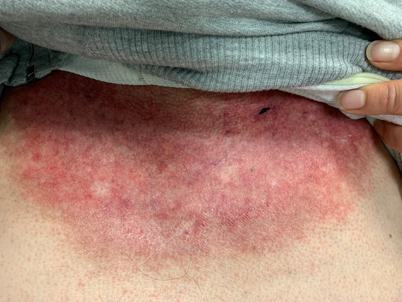
Anti-TIF1γ Dermatomyositis with Scleroderma-Like Pattern
A 67-year-old man with chronic anti-TIF1γ-positive dermatomyositis and a scleroderma-like capillaroscopy pattern.
Clinical context: Two-year history of V-neck lesions and hypomyopathic weakness on chronic low-dose methotrexate and prednisone.
Capillaroscopy: Capillaroscopy shows severely reduced capillary density (2.7/mm), architectural disorganization, frequent dilations, giant capillaries, and abnormal/ramified forms (2/3 on the semi-quantitative scale), consistent with a scleroderma-like pattern.
Clinical images
Key Capillaroscopic Findings:
- • Severe capillary loss (2.7/mm)
- • Giant capillaries (36.7%) and frequent dilations (41.1%)
- • Abnormal or ramified capillaries (37.8%)
- • Architectural disorganization
Juvenile Dermatomyositis with Gottron Lesions and a Scleroderma-Like Pattern
A 13-year-old girl with Gottron lesions, no muscle weakness, normal CPK, and a markedly pathological capillaroscopy at disease onset.
Clinical context: The patient was referred to rheumatology because of skin lesions compatible with juvenile dermatomyositis. Examination showed Gottron papules on the fingers and Gottron signs on the ankles, elbows, and knees. She had no muscle weakness, with an MMT score of 80/80. Laboratory testing showed normal CPK, positive ANA, and weak anti-PM/Scl positivity, with the remaining autoantibodies negative. Treatment was started with hydroxychloroquine and intravenous immunoglobulin.
Capillaroscopy: The baseline capillaroscopy is compatible with juvenile dermatomyositis and shows a severe scleroderma-like microangiopathy, with marked capillary loss, frequent enlarged and giant capillaries, hemorrhages, abnormal or arborizing shapes, and architectural disorganization.
Key Capillaroscopic Findings:
- • Marked capillary loss (4.46/mm)
- • Frequent giant capillaries (21.1%) and enlarged capillaries (64.8%)
- • Hemorrhages and abnormal/arborizing capillaries in an active myositis-associated pattern
Juvenile Dermatomyositis anti-NXP2 with Capillaroscopic Improvement
A 12-year-old girl with muscle weakness, markedly elevated CPK, and no skin disease, followed with two capillaroscopies during treatment response.
Clinical context: The patient presented with several weeks of shoulder and pelvic girdle weakness, without skin involvement or other symptoms. Laboratory testing showed CPK elevation up to 8000 U/L, lymphopenia, thrombocytopenia, and low-titer positive ANA. Whole-body MRI showed muscle edema in the girdles. The initial myositis panel was negative, so the early scleroderma-like capillaroscopy pattern helped support the diagnosis of juvenile dermatomyositis despite the absence of skin disease or specific autoantibodies. An extended panel later detected anti-NXP2 antibodies. Treatment was started with hydroxychloroquine, mycophenolate mofetil, and intravenous immunoglobulin, followed by clinical improvement and CK reduction.
Capillaroscopy: The June 2024 capillaroscopy showed an early scleroderma-like pattern compatible with juvenile dermatomyositis, including reduced density, frequent enlarged capillaries, sparse giant capillaries, and hemorrhages. The December 2024 follow-up showed disappearance of giant capillaries and evolution toward a non-specific pattern, with improved capillary density.
Key Capillaroscopic Findings:
- • Initial reduced density (5.83/mm) with frequent enlarged capillaries (68.8%)
- • Initial sparse giant capillaries (2.6%) and hemorrhages
- • Follow-up density improved to 6.72/mm, with disappearance of giant capillaries
Juvenile Dermatomyositis anti-TIF1 with Active Scleroderma-Like Pattern
A 12-year-old girl with Gottron lesions, facial erythema, muscle weakness, dysphagia, and anti-TIF1 antibodies.
Clinical context: The patient was referred for skin lesions, including Gottron lesions and facial erythema, together with muscle weakness and dysphagia. Complementary tests were compatible with juvenile dermatomyositis, and anti-TIF1 antibodies were positive.
Capillaroscopy: Capillaroscopy shows a severe active scleroderma-like pattern, with very low capillary density, numerous giant capillaries, frequent enlarged capillaries, arborizing abnormal shapes, and architectural disorganization. This morphology is highly suggestive of active microangiopathy in juvenile dermatomyositis.
Key Capillaroscopic Findings:
- • Severe capillary loss (2.89/mm)
- • Very frequent giant capillaries (44.6%)
- • Abnormal/arborizing capillaries and marked architectural disorganization
Mixed Connective Tissue Disease Presenting with Raynaud Phenomenon
A 10-year-old girl with Raynaud phenomenon, puffy fingers, anti-RNP positivity, and later carpal arthritis.
Clinical context: The patient presented with a 3-month history of Raynaud phenomenon. Her family history included a father with systemic lupus erythematosus. Examination showed swelling of the fingers and delayed capillary refill. Laboratory testing showed anti-RNP positivity. Months later she developed carpal arthritis, leading to a diagnosis of mixed connective tissue disease.
Capillaroscopy: The initial capillaroscopy showed an early scleroderma pattern, supporting the diagnosis in the appropriate clinical and serological context. On follow-up, clinical improvement was accompanied by improvement of capillaroscopic findings, with higher density, disappearance of giant capillaries, and evolution toward a non-specific pattern.
Key Capillaroscopic Findings:
- • Initial scleroderma pattern with reduced density (6.06/mm)
- • Initial enlarged capillaries (70.3%), sparse giant capillaries, and hemorrhages
- • Follow-up improvement to 7.46/mm density, without giant capillaries