
Rheumatology Research Lead, Sura Biosciences Division
Dr. Miguel Antonio Mesa is a rheumatologist and internist, currently working as the rheumatology research line lead at Sura Biosciences Division. He also leads the microcirculation study group at the Colombian Association of Rheumatology. His academic interests focus on capillaroscopy and the use of advanced therapies in autoimmune diseases.
Contributed cases and studies (4)
Normal Pattern: Representative Case
Complete capillaroscopy study with preserved density, regular morphology, and no scleroderma-pattern features.
Systemic Sclerosis with anti-U3 RNP: Late Microangiopathy
Patient with systemic sclerosis, anti-U3 RNP antibodies, and severe microvascular disease with features of the late Cutolo pattern.
Anticentromere-Positive Systemic Sclerosis with Inter-Digit Pattern Heterogeneity
A 61-year-old Latin American woman with long-standing Raynaud phenomenon and recently diagnosed anticentromere-positive systemic sclerosis, showing markedly different capillaroscopic patterns across digits.
Mixed Connective Tissue Disease with an Unclassifiable Scleroderma-Like Pattern
A patient with mixed connective tissue disease, strongly positive anti-RNP antibodies, and a scleroderma-like capillaroscopic pattern featuring megacapillaries and arborizing forms.
Images in the atlas (24)
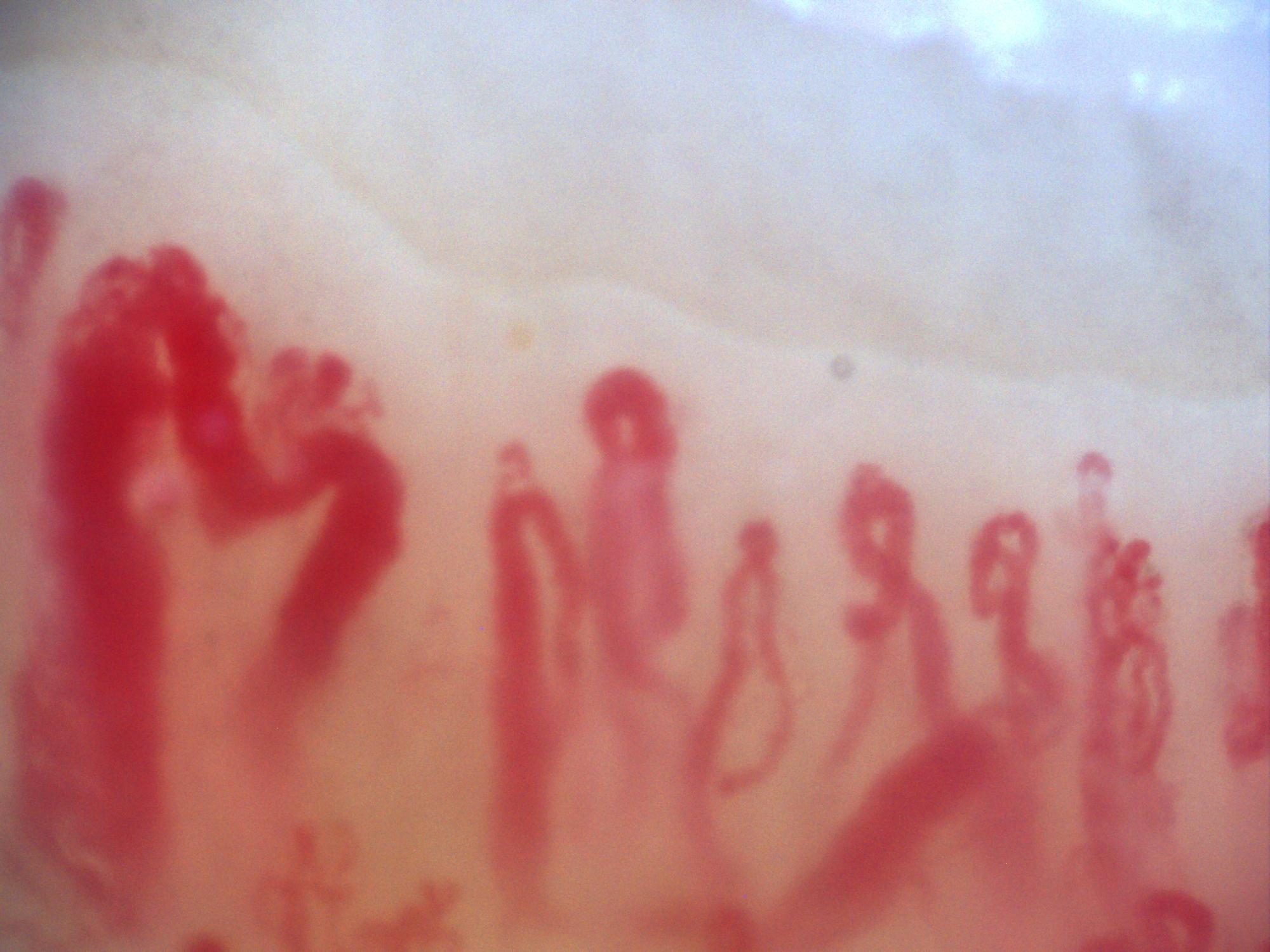
200x capillaroscopy image shared by Miguel Antonio Mesa Navas as an example of an active background with prominent megacapillaries.
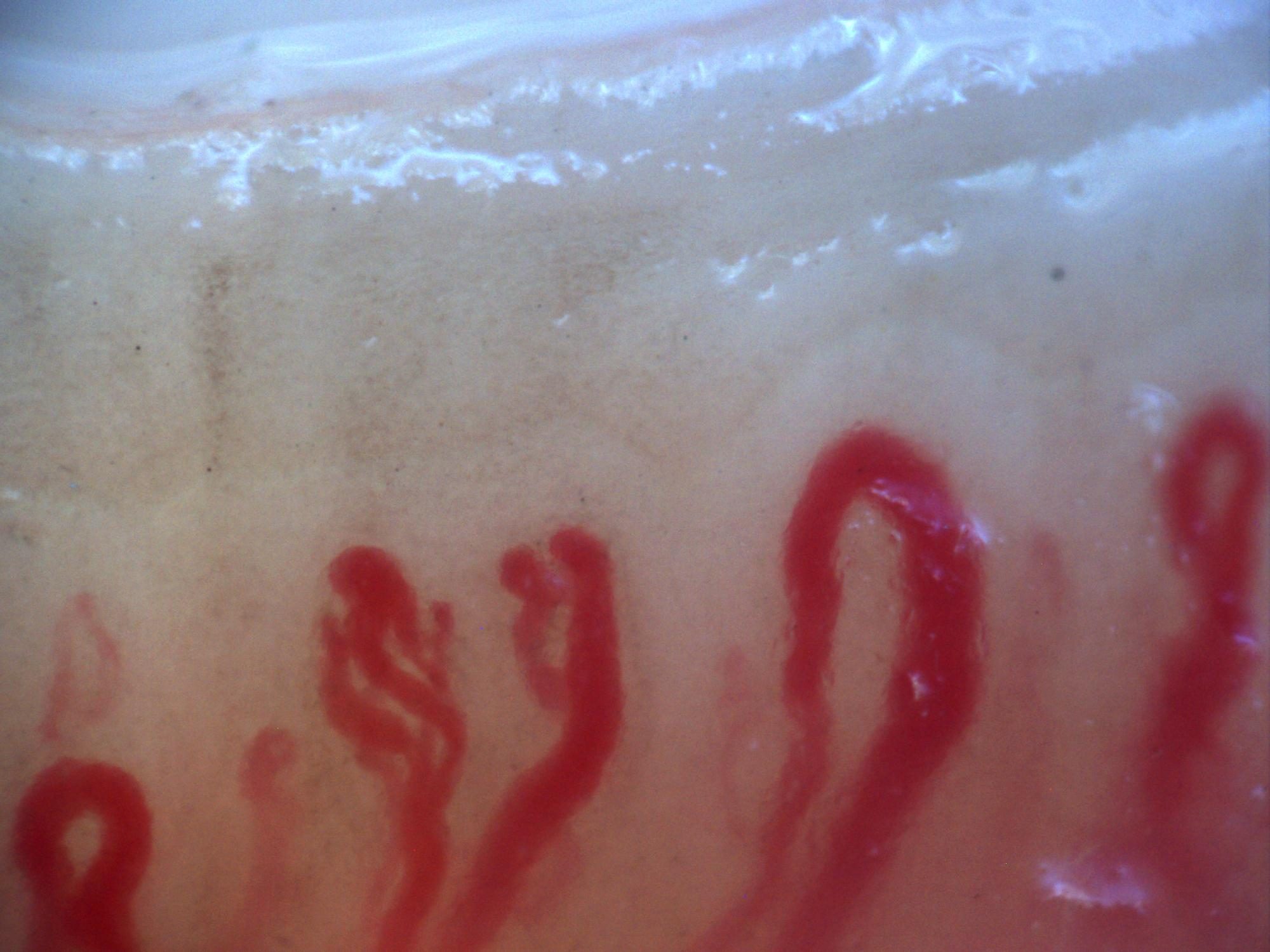
Another 200x capillaroscopy image shared by Miguel Antonio Mesa Navas as an example of an active background with prominent megacapillaries.
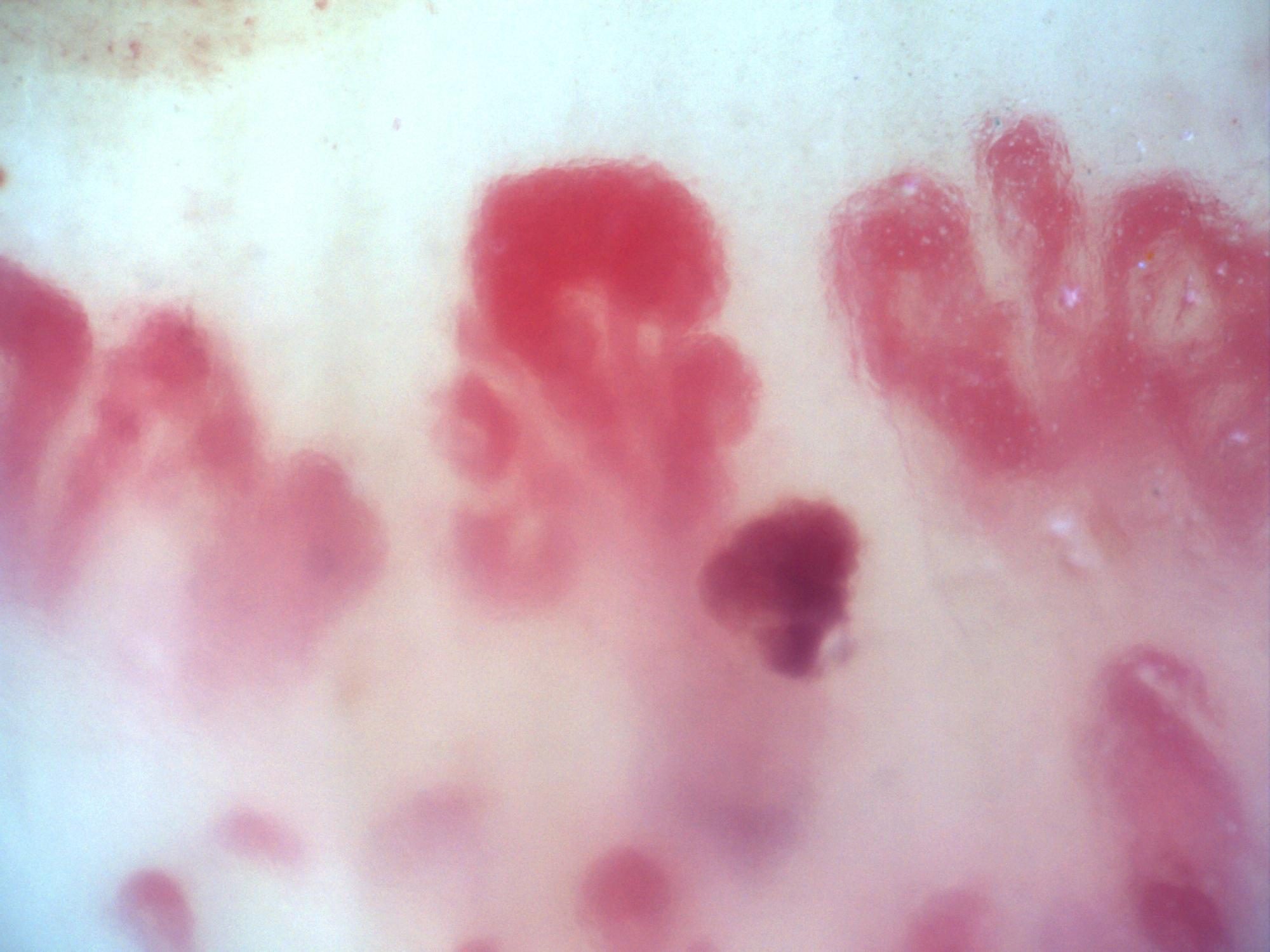
200x capillaroscopy image shared as an additional example of megacapillaries on an active background.
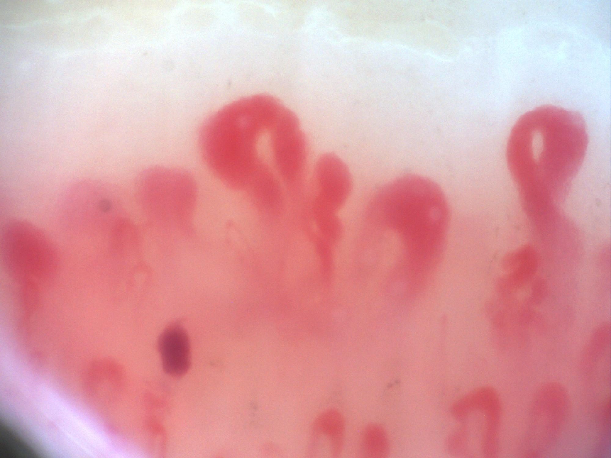
200x capillaroscopy image sent by Miguel Antonio Mesa Navas within the series of megacapillary examples.
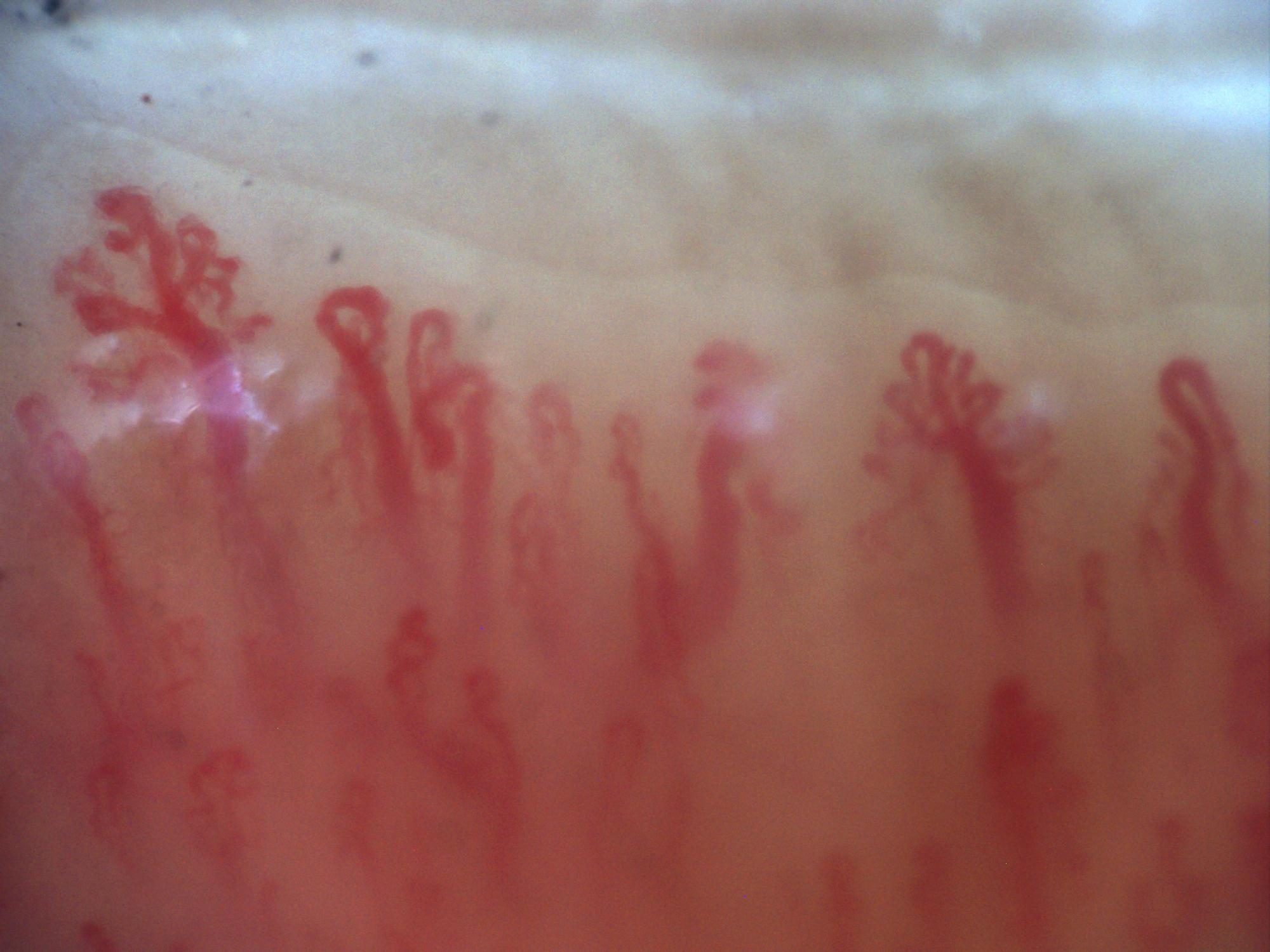
200x capillaroscopy image with a branching capillary on the left side of the field.
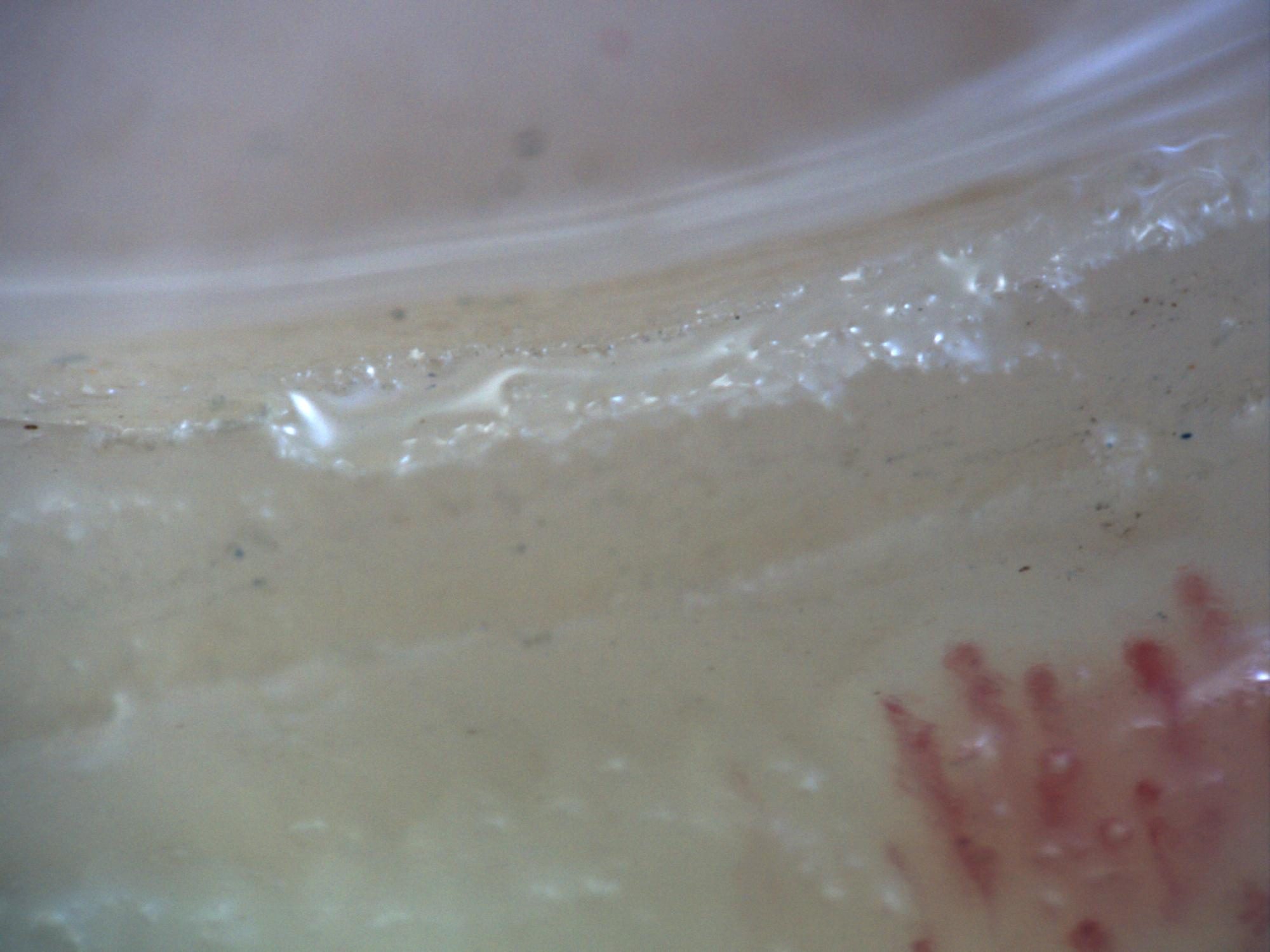
200x capillaroscopy image showing excessive pressure on the nail, with disappearance of the visible capillary bed along the left margin.
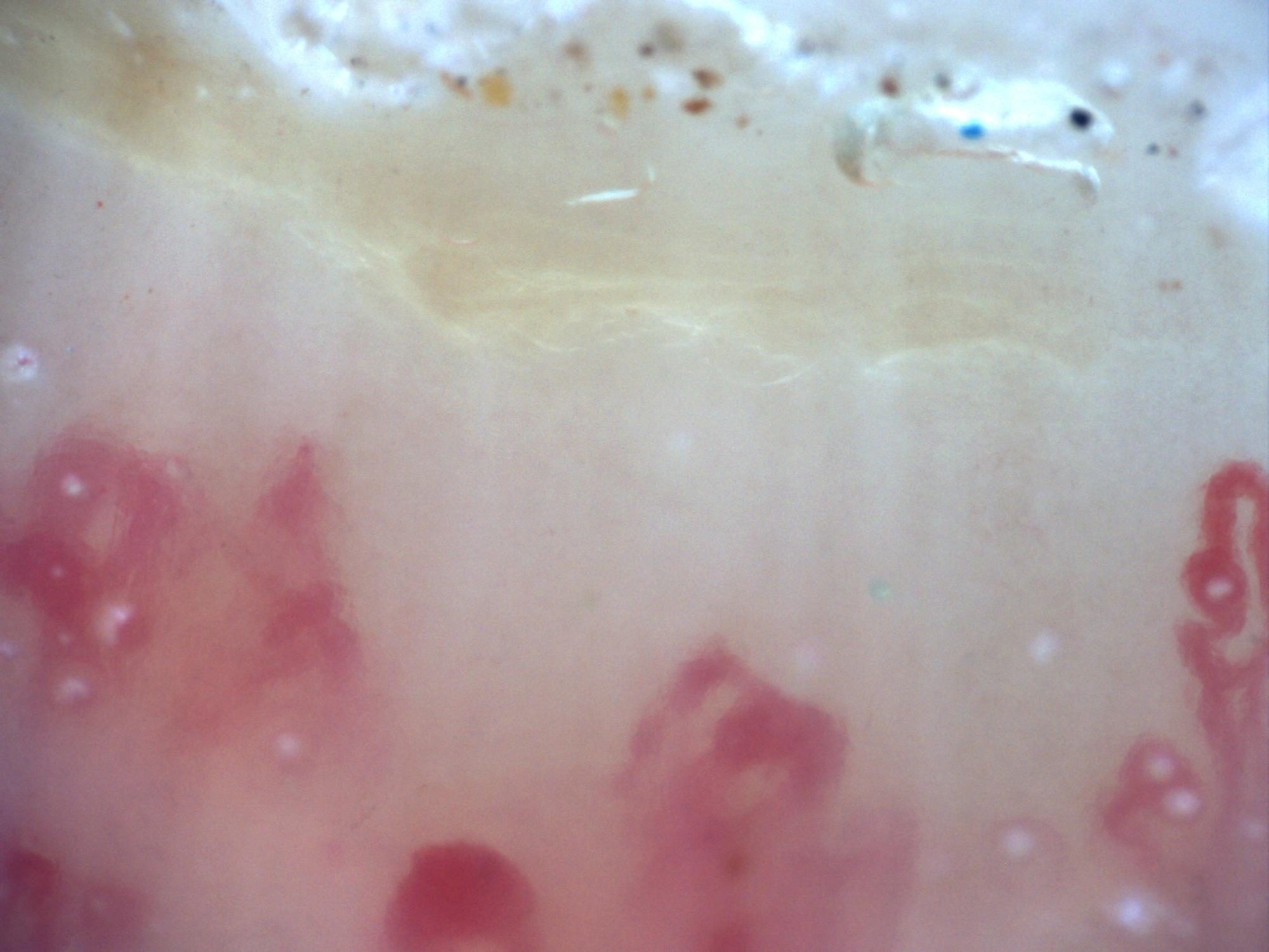
200x capillaroscopy image showing an extensive capillary-free space compatible with an avascular area.
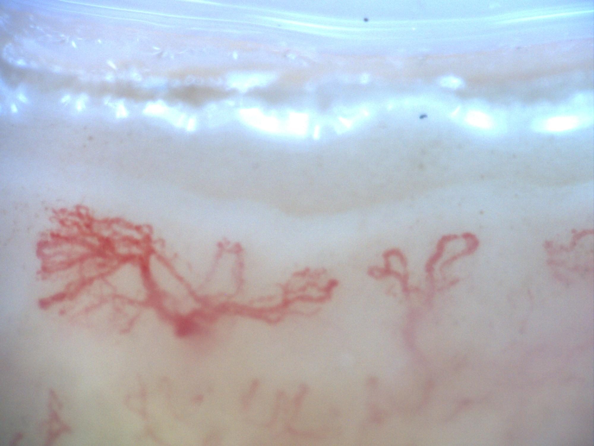
200x capillaroscopy image from a patient with systemic sclerosis showing a late pattern with marked disorganization and neoangiogenesis, without visible megacapillaries.
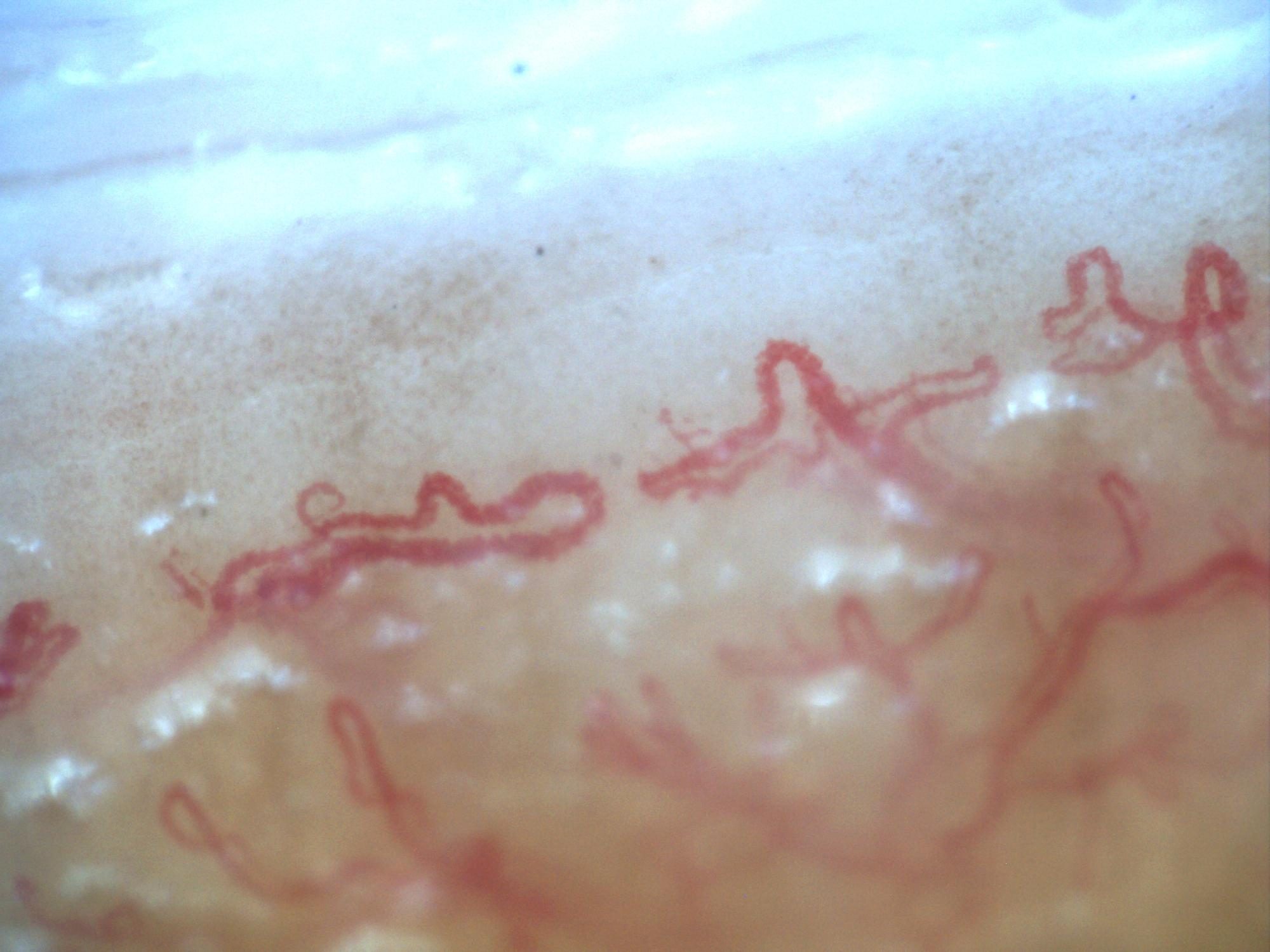
Second 200x capillaroscopy image from systemic sclerosis with a late pattern, marked disorganization, and neoangiogenesis.
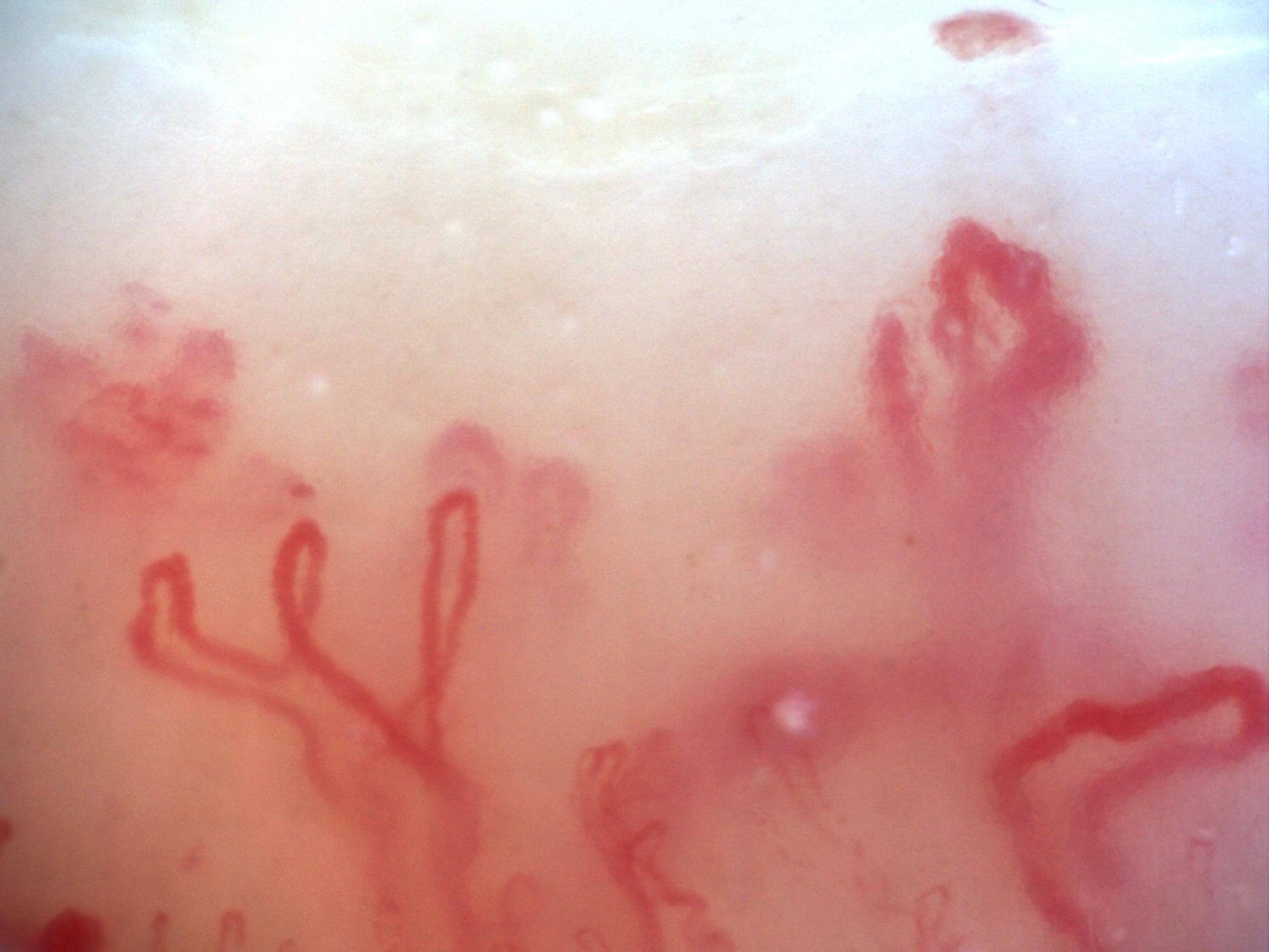
200x capillaroscopy image documenting pericapillary edema on an active background, visible as a fog-like haze between capillaries.
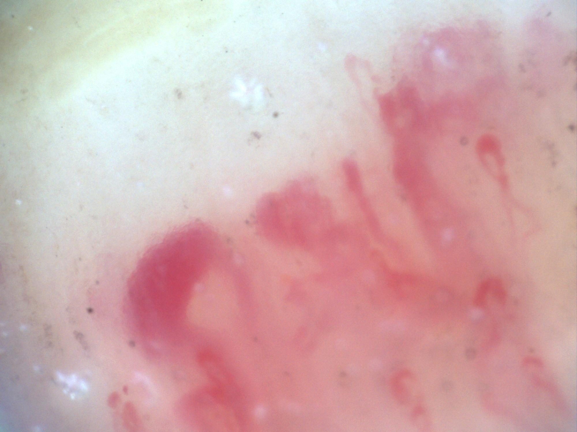
200x capillaroscopy image from systemic sclerosis showing megacapillaries and a ground-glass stromal pattern described by the contributor as reflecting fibrosis, with some associated pericapillary edema.
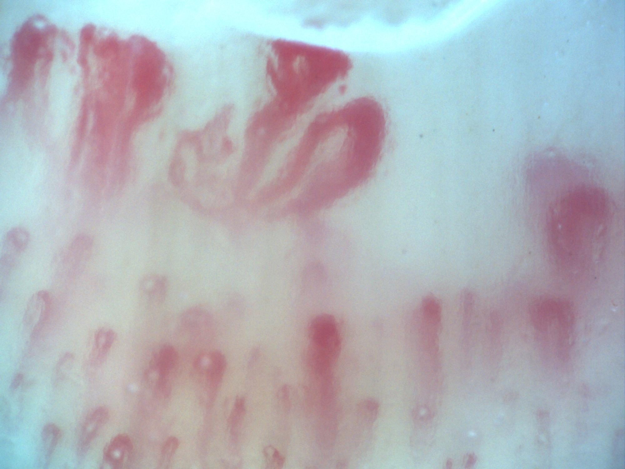
Second 200x capillaroscopy image from systemic sclerosis showing megacapillaries, pericapillary edema, and a ground-glass stromal appearance.
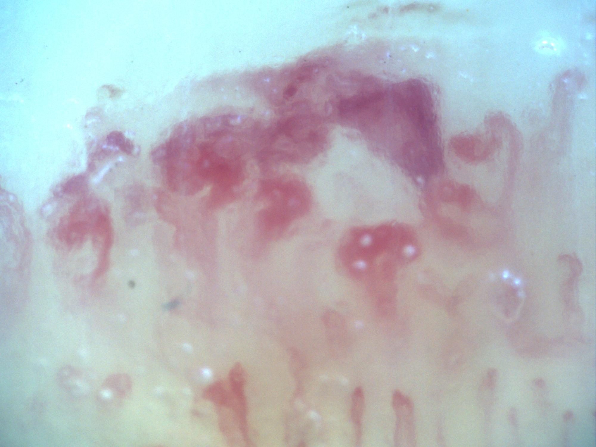
200x capillaroscopy image from systemic sclerosis showing a microvascular hemorrhage with the typical smoke-plume appearance.
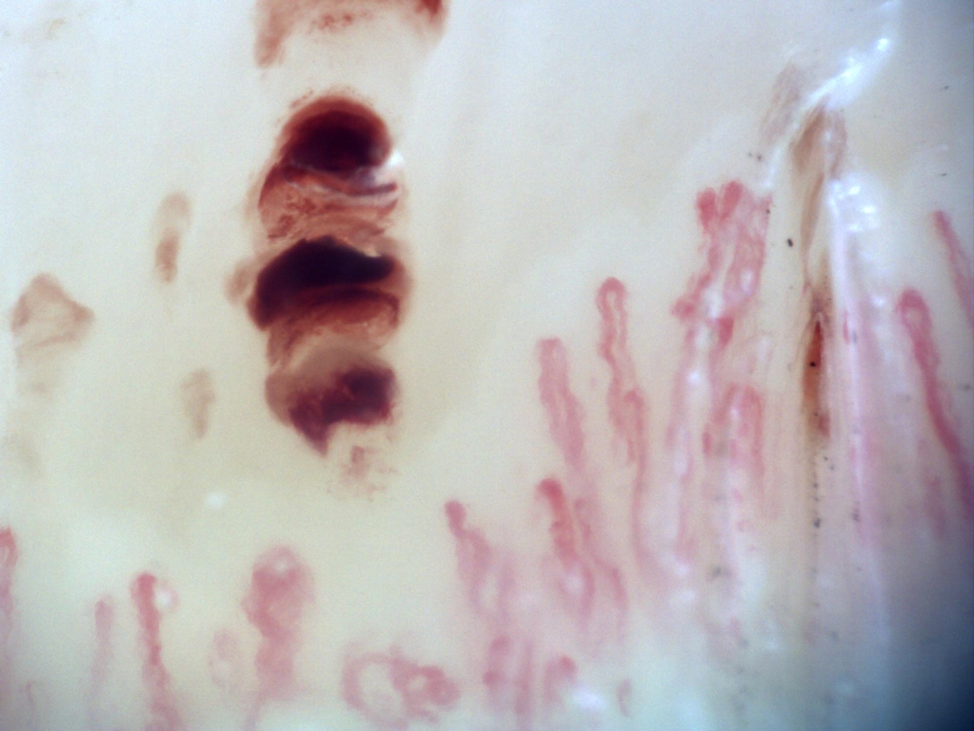
200x capillaroscopy image from systemic sclerosis showing a representative microvascular hemorrhagic lesion.
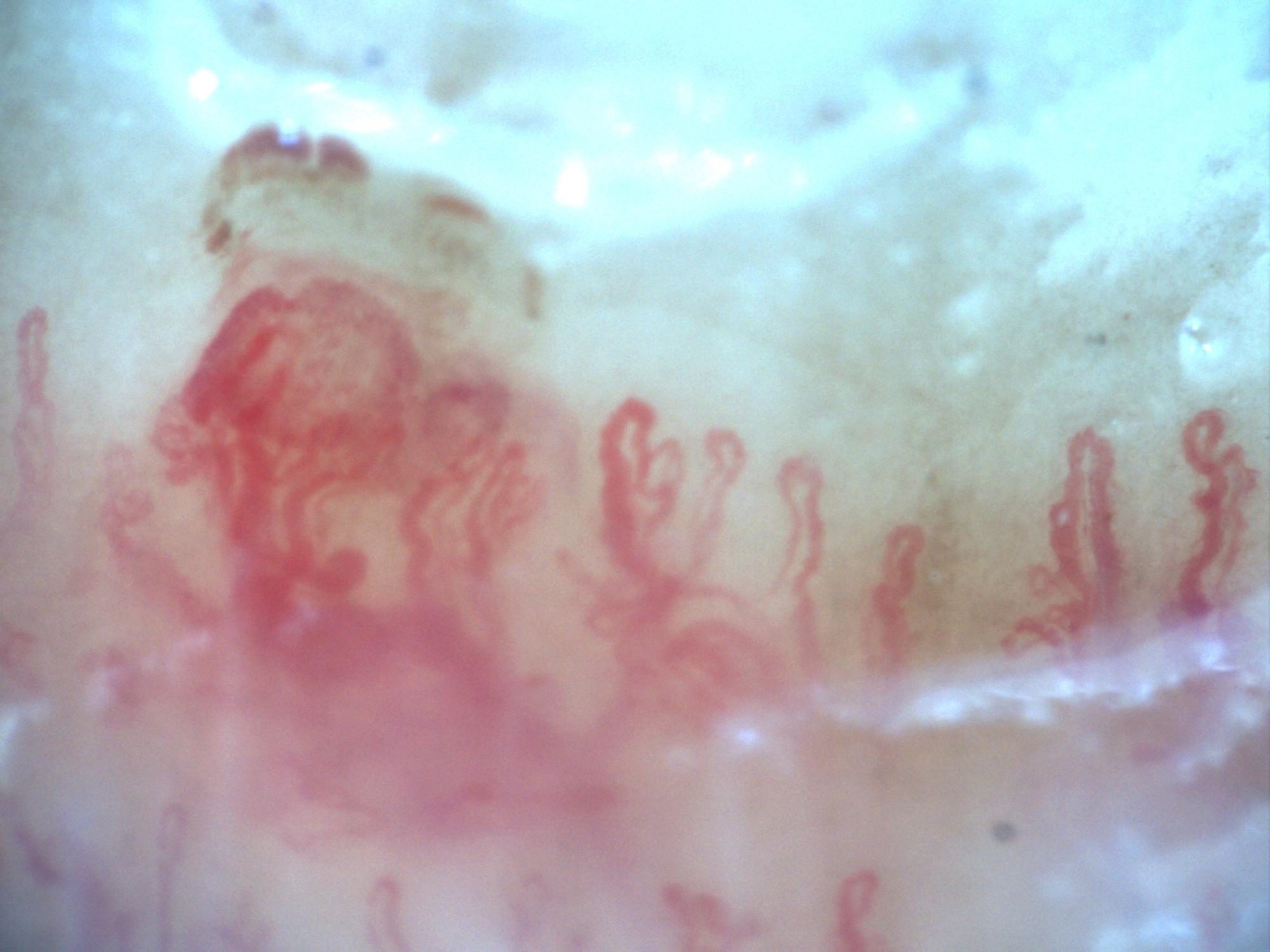
200x capillaroscopy image capturing a hemorrhage at the moment of its formation.
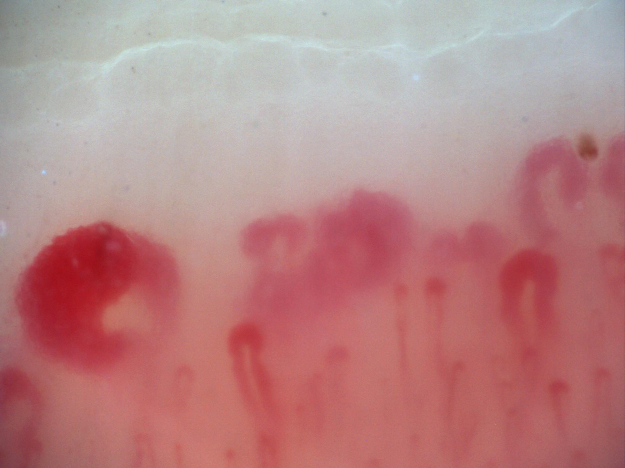
200x capillaroscopy image from systemic sclerosis showing a very large megacapillary.
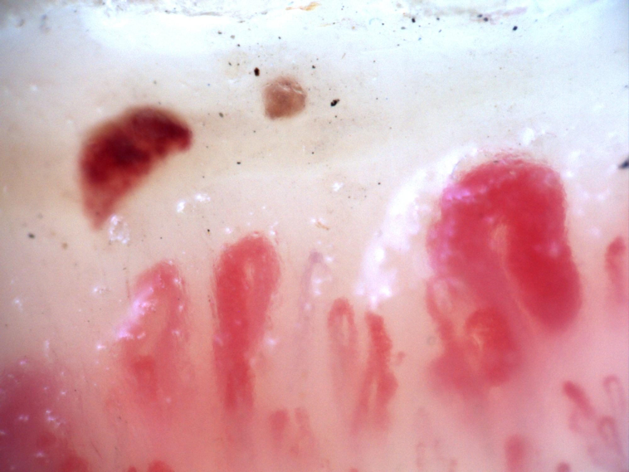
Second 200x capillaroscopy image with a large megacapillary in systemic sclerosis.
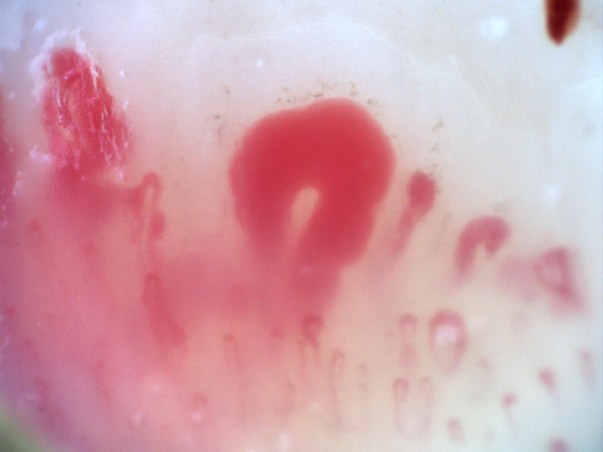
Third 200x capillaroscopy image showing large megacapillaries in a systemic sclerosis setting.
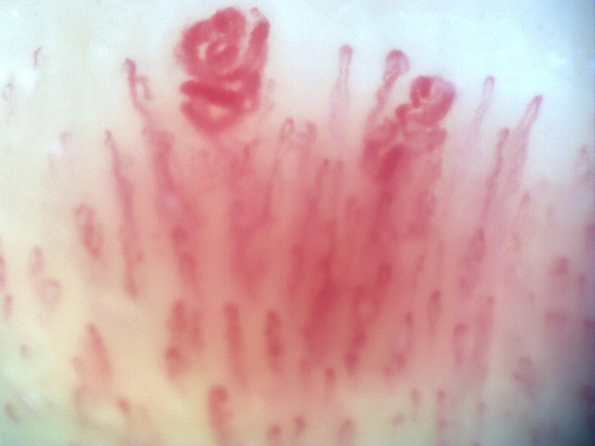
200x capillaroscopy image showing a giant capillary with a coiled or ball-of-yarn appearance.
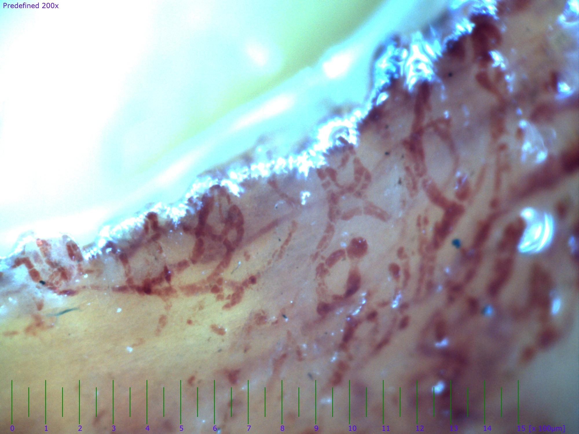
Unusual capillaroscopy image contributed as a personal reference case, showing widespread capillary thrombosis in a critically ischemic digit. The contributor highlighted the rarity of this finding and recommended clinicopathologic correlation.
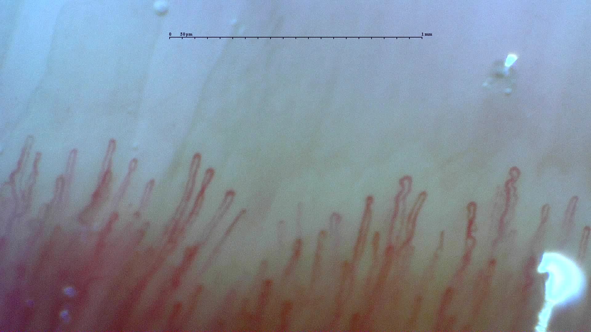 Normal reference: distal capillary row
A field from the normal reference study, showing the distal row of narrow loops. Use the full study to compare density and shape across fingers.
View the complete study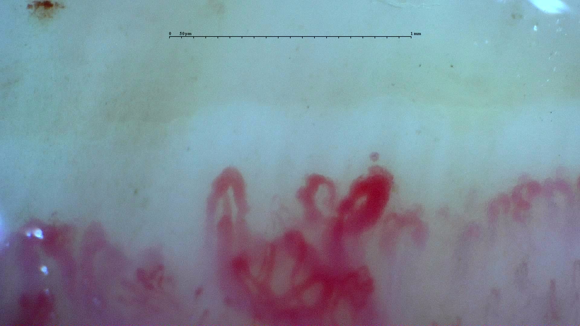 Anti-U3 RNP systemic sclerosis: irregular loops
Irregular enlarged loops in the anti-U3 RNP case. Although giants remain present, the final report classifies the overall microangiopathy as late.
View the complete study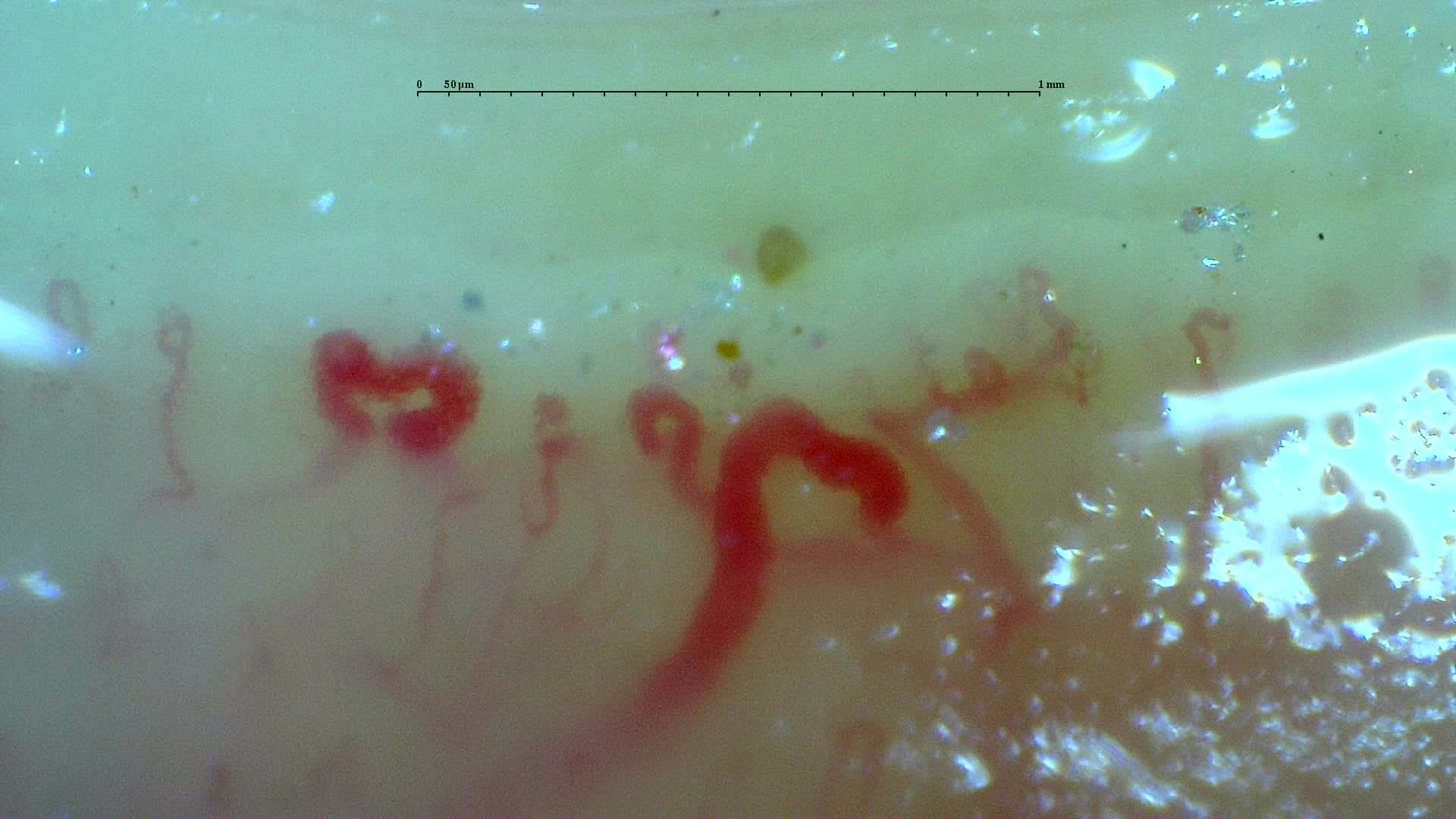 Inter-digit heterogeneity: selected abnormal field
A disorganized field with enlarged loops and surface reflections from the inter-digit heterogeneity case. Other fields differ markedly; inspect the full study and its image-quality caveats.
View the complete study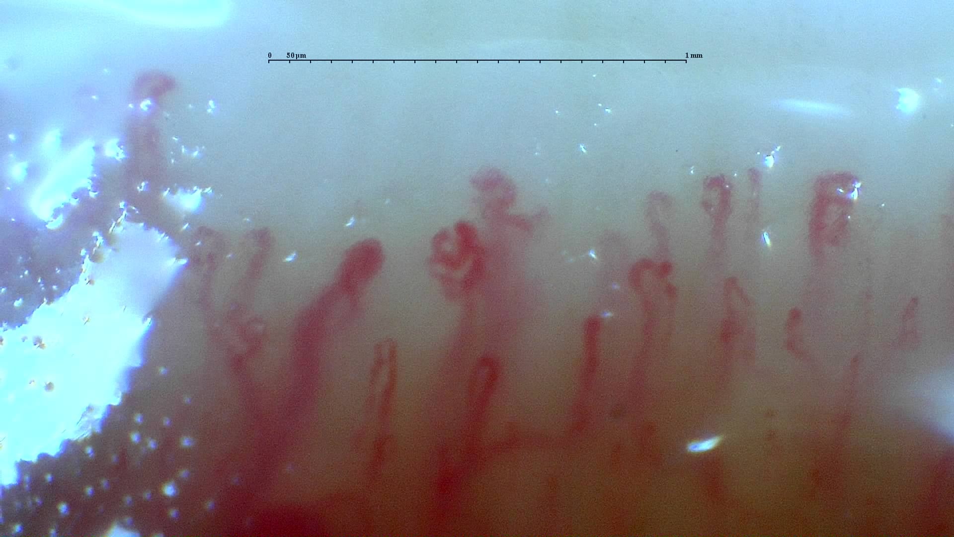 Mixed connective tissue disease: irregular branching forms
Irregular and branching forms in the anti-RNP-positive mixed connective tissue disease case. The source report describes a scleroderma-like pattern without an assignable Cutolo subtype.
View the complete study