Sample image
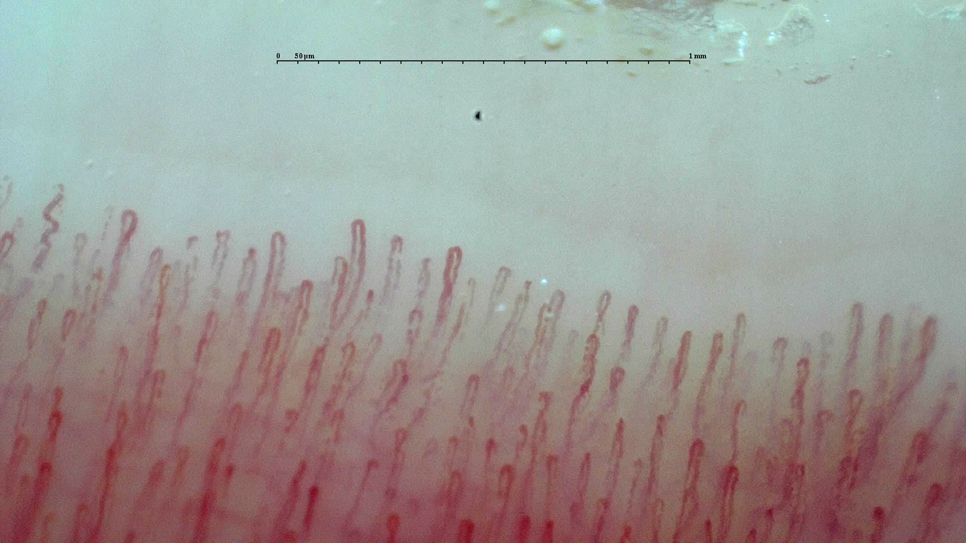
Normal distal row
Observe orderly capillary loops, preserved density, and no scleroderma-pattern features.
Learn nailfold capillaroscopy with real clinical images
A practical visual resource for learning nailfold capillaroscopy, interpreting patterns, and exploring complete clinical cases.
Nailfold capillaroscopy lets you examine nailfold capillaries non-invasively. It is especially useful for assessing microvascular involvement in connective tissue diseases, particularly systemic sclerosis.
Use this site to review the fundamentals, compare patterns, study clinical cases, and open complete studies so morphology, density, and the distribution of findings stay connected.
Before getting into classifications, look at what the findings actually look like. The site combines real images and teaching figures to help you recognize morphology, density, artifacts, and patterns.
Sample image
Observe orderly capillary loops, preserved density, and no scleroderma-pattern features.
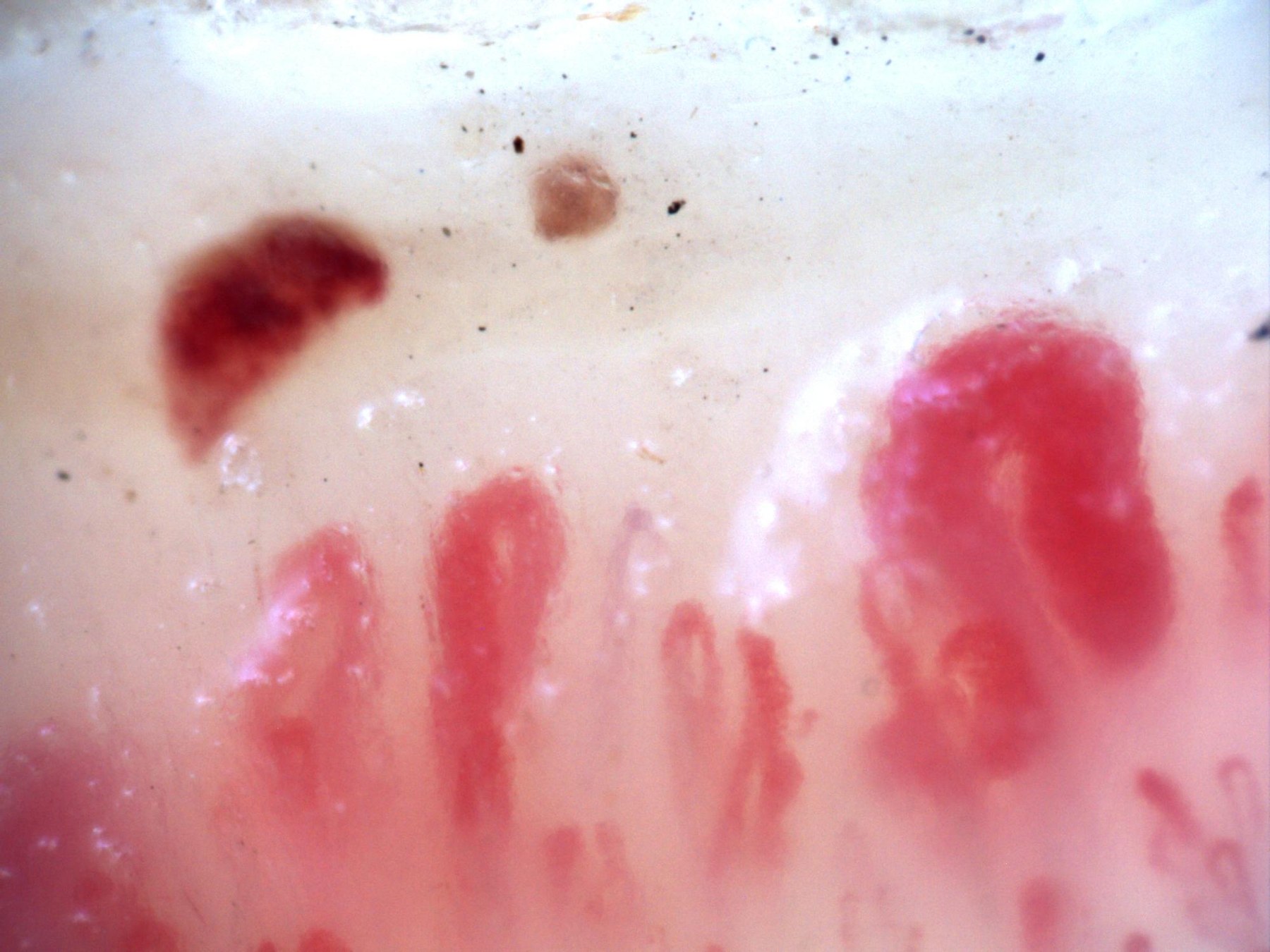
Sample image
Recognize giant capillaries, hemorrhages, and architectural changes in systemic-sclerosis-type microangiopathy.
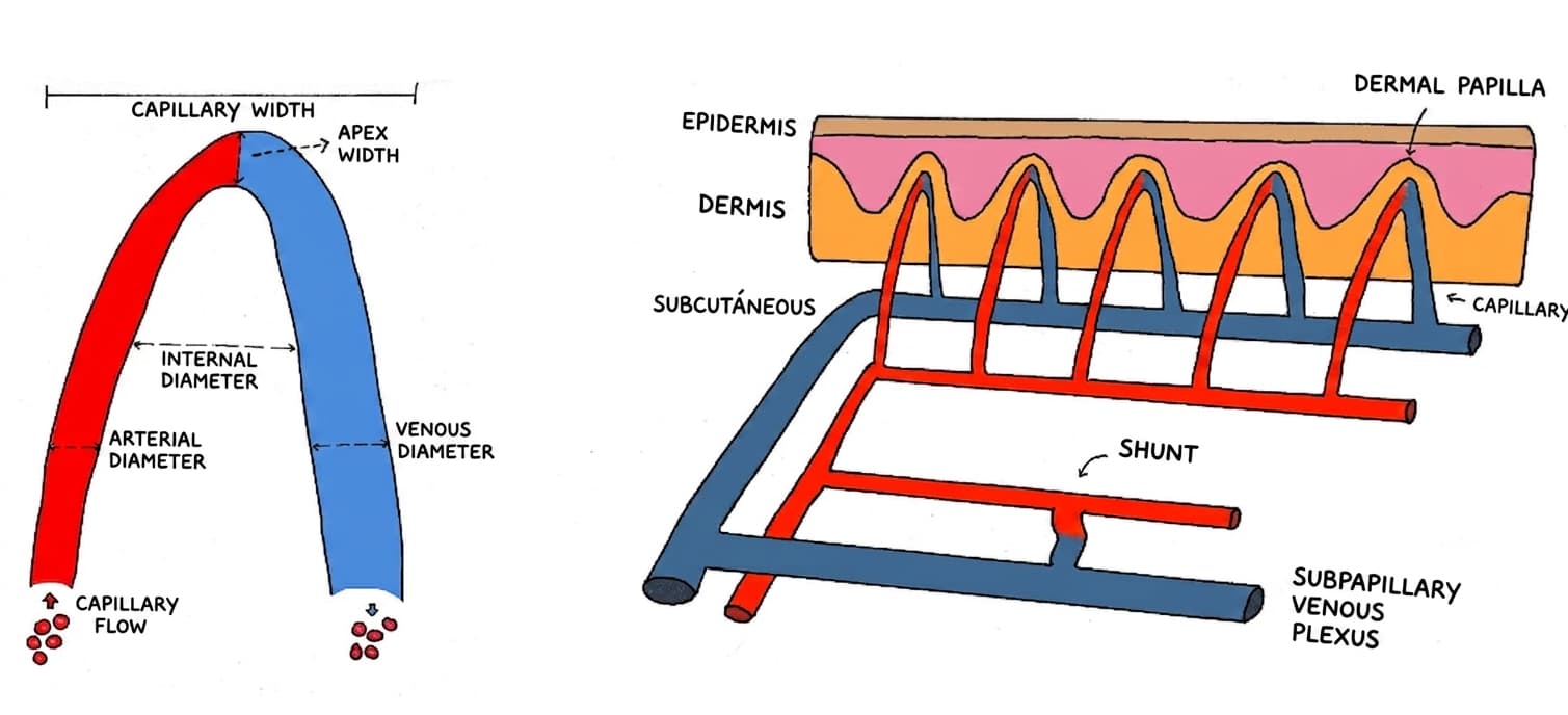
Figure
Locate the distal row and understand why the nailfold is a useful window into the microcirculation.
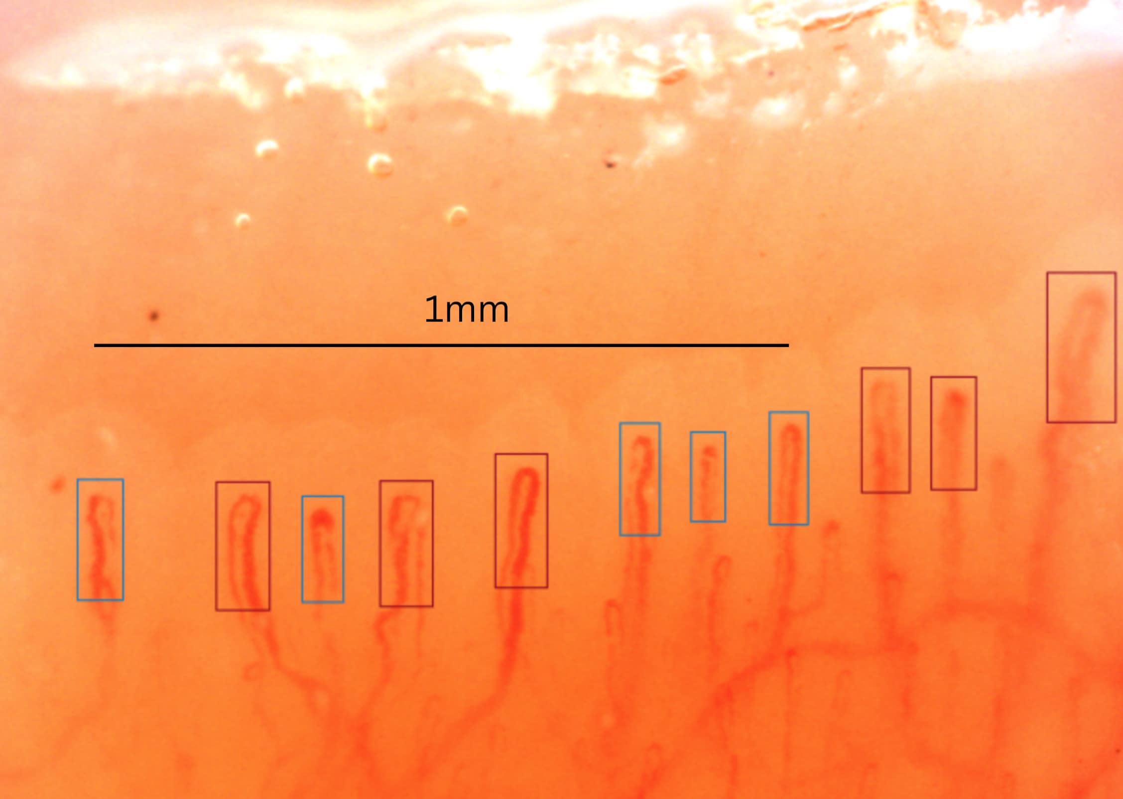
Figure
Learn how to count visible capillaries and translate findings into a reproducible score.
Interactive viewer
On the clinical cases and patterns pages, you can open complete studies in the embedded Capillary.io viewer. Move through fields and fingers, compare images, and keep the visual pattern connected to the quantitative data.
Representative active systemic sclerosis pattern study.
Guillén Del Castillo A, Lledó-Ibáñez GM, Sáez Comet L, Freire Dapena M, Mesa Navas M, Martín Cascón M, et al. Med Clin (Barc). 2026;166(6):107426. doi: 10.1016/j.medcli.2026.107426. PMID: 42013567.
Large multicenter study showing that all non-thumb fingers and four nailbed areas should be assessed for reliable systemic sclerosis pattern classification.
Mugii N, Hamaguchi Y, Fushida N, Fujii K, Nishio J, Kudo K, et al. J Dermatol. 2026 Feb;53(2):318-322. PMID: 41312673.
Shows that nailfold videocapillaroscopy findings vary by anti-ARS antibody subtype in antisynthetase syndrome, with anti-KS associated with milder microvascular involvement.
Ingegnoli F, Pireddu D, Platania E, De Angelis R, Alunno A, Ariani A, et al. Clin Exp Rheumatol. 2026 Jan 15. doi: 10.55563/clinexprheumatol/2z4j95. PMID: 41537537.
Delphi-based reporting guidance defining 23 nailfold videocapillaroscopy report items, including 16 mandatory items for routine clinical practice.
Boulon C, Larrouture I, Blaise S, Mangin M, Chevoir JD, Senet P, et al. Microvasc Res. 2026 Jan;163:104874. PMID: 40967366.
Prospective SCLEROCAP follow-up showing that capillaroscopy can help identify patients with systemic sclerosis at higher risk of severe 3-year progression.
Bilancini S, Lucchi M, Trevisan G, Di Pino L, Tucci S. Acta Dermatovenerol Alp Pannonica Adriat. 2025 Dec;34(4):177-180. PMID: 41420618.
Shows that 45.3% of patients with acrocyanosis had a secondary form, most often associated with connective tissue disease among secondary cases.
Yayla ME, Aydın A, Kılıçaslan M, Kalkan M, Güzel MS, Shikhaliyeva A, et al. Diagnostics (Basel). 2025 Nov 18;15(22):2912. PMID: 41300936.
Reports high classification performance from deep-learning models for videocapillaroscopy images, supporting the current move toward AI-assisted interpretation.
Bhandare P, Naik R, Mahesh Y, Ghodge R. Indian Dermatol Online J. 2025 Nov 1;16(6):928-933. PMID: 40814970.
Cross-sectional vasculitis study linking capillary disorganization, dropouts, receding capillaries, and avascular areas with systemic involvement and ulcerative lesions.